 Movie
Movie Controller


+ Open data

- Basic information
| Entry | Database: PDB / ID: 7za3 | ||||||
|---|---|---|---|---|---|---|---|
| Title | GPC3-Unc5D octamer structure and role in cell migration | ||||||
 Components Components |
| ||||||
 Keywords Keywords | SIGNALING PROTEIN / Complex / Cell migration / Glycan-Glycan Interaction | ||||||
| Function / homology |  Function and homology information Function and homology informationGlycosaminoglycan-protein linkage region biosynthesis / HS-GAG biosynthesis / HS-GAG degradation / peptidyl-dipeptidase inhibitor activity / mesonephric duct morphogenesis / body morphogenesis / cell proliferation involved in kidney development / netrin receptor activity / regulation of non-canonical Wnt signaling pathway / Retinoid metabolism and transport ...Glycosaminoglycan-protein linkage region biosynthesis / HS-GAG biosynthesis / HS-GAG degradation / peptidyl-dipeptidase inhibitor activity / mesonephric duct morphogenesis / body morphogenesis / cell proliferation involved in kidney development / netrin receptor activity / regulation of non-canonical Wnt signaling pathway / Retinoid metabolism and transport / mesenchymal cell proliferation involved in ureteric bud development / cell proliferation involved in metanephros development / cell migration involved in gastrulation / Regulation of Insulin-like Growth Factor (IGF) transport and uptake by Insulin-like Growth Factor Binding Proteins (IGFBPs) / Post-translational protein phosphorylation / negative regulation of growth / : / positive regulation of Wnt signaling pathway, planar cell polarity pathway / positive regulation of BMP signaling pathway / coronary vasculature development / regulation of neuron migration / positive regulation of smoothened signaling pathway / regulation of growth / embryonic hindlimb morphogenesis / regulation of canonical Wnt signaling pathway / pyramidal neuron differentiation / anterior/posterior axis specification / Wnt signaling pathway, planar cell polarity pathway / branching involved in ureteric bud morphogenesis / smoothened signaling pathway / bone mineralization / positive regulation of endocytosis / canonical Wnt signaling pathway / side of membrane / lung development / axon guidance / osteoclast differentiation / animal organ morphogenesis / epithelial cell proliferation / positive regulation of D-glucose import / negative regulation of smoothened signaling pathway / response to bacterium / kidney development / negative regulation of canonical Wnt signaling pathway / Golgi lumen / negative regulation of epithelial cell proliferation / positive regulation of protein catabolic process / positive regulation of canonical Wnt signaling pathway / lysosome / negative regulation of cell population proliferation / apoptotic process / cell surface / plasma membrane Similarity search - Function | ||||||
| Biological species |  | ||||||
| Method |  X-RAY DIFFRACTION / SYNCHROTRON / MOLECULAR REPLACEMENT / Resolution: 4 Å X-RAY DIFFRACTION / SYNCHROTRON / MOLECULAR REPLACEMENT / Resolution: 4 Å | ||||||
 Authors Authors | Akkermans, O. / Delloye-Bourgeois, C. / Peregrina, C. / Carrasquero, M. / Kokolaki, M. / Berbeira-Santana, M. / Chavent, M. / Reynaud, F. / Ritu, R. / Agirre, J. ...Akkermans, O. / Delloye-Bourgeois, C. / Peregrina, C. / Carrasquero, M. / Kokolaki, M. / Berbeira-Santana, M. / Chavent, M. / Reynaud, F. / Ritu, R. / Agirre, J. / Aksu, M. / White, E. / Lowe, E. / Ben Amar, D. / Zaballa, S. / Huo, J. / Pakos, I. / McCubbin, P. / Comoletti, D. / Owens, R. / Robinson, C. / Castellani, V. / del Toro, D. / Seiradake, E. | ||||||
| Funding support |  United Kingdom, 1items United Kingdom, 1items
| ||||||
 Citation Citation | Journal: Cell / Year: 2022 Title: GPC3-Unc5 receptor complex structure and role in cell migration. Authors: Akkermans, O. / Delloye-Bourgeois, C. / Peregrina, C. / Carrasquero-Ordaz, M. / Kokolaki, M. / Berbeira-Santana, M. / Chavent, M. / Reynaud, F. / Raj, R. / Agirre, J. / Aksu, M. / White, E.S. ...Authors: Akkermans, O. / Delloye-Bourgeois, C. / Peregrina, C. / Carrasquero-Ordaz, M. / Kokolaki, M. / Berbeira-Santana, M. / Chavent, M. / Reynaud, F. / Raj, R. / Agirre, J. / Aksu, M. / White, E.S. / Lowe, E. / Ben Amar, D. / Zaballa, S. / Huo, J. / Pakos, I. / McCubbin, P.T.N. / Comoletti, D. / Owens, R.J. / Robinson, C.V. / Castellani, V. / Del Toro, D. / Seiradake, E. #1: Journal: Acta Crystallogr., Sect. D: Biol. Crystallogr. / Year: 2012Title: Towards automated crystallographic structure refinement with phenix.refine. Authors: Afonine, P.V. #2: Journal: Acta Crystallogr D Struct Biol / Year: 2019 Title: Macromolecular structure determination using X-rays, neutrons and electrons: recent developments in Phenix. Authors: Dorothee Liebschner / Pavel V Afonine / Matthew L Baker / Gábor Bunkóczi / Vincent B Chen / Tristan I Croll / Bradley Hintze / Li Wei Hung / Swati Jain / Airlie J McCoy / Nigel W Moriarty ...Authors: Dorothee Liebschner / Pavel V Afonine / Matthew L Baker / Gábor Bunkóczi / Vincent B Chen / Tristan I Croll / Bradley Hintze / Li Wei Hung / Swati Jain / Airlie J McCoy / Nigel W Moriarty / Robert D Oeffner / Billy K Poon / Michael G Prisant / Randy J Read / Jane S Richardson / David C Richardson / Massimo D Sammito / Oleg V Sobolev / Duncan H Stockwell / Thomas C Terwilliger / Alexandre G Urzhumtsev / Lizbeth L Videau / Christopher J Williams / Paul D Adams /   Abstract: Diffraction (X-ray, neutron and electron) and electron cryo-microscopy are powerful methods to determine three-dimensional macromolecular structures, which are required to understand biological ...Diffraction (X-ray, neutron and electron) and electron cryo-microscopy are powerful methods to determine three-dimensional macromolecular structures, which are required to understand biological processes and to develop new therapeutics against diseases. The overall structure-solution workflow is similar for these techniques, but nuances exist because the properties of the reduced experimental data are different. Software tools for structure determination should therefore be tailored for each method. Phenix is a comprehensive software package for macromolecular structure determination that handles data from any of these techniques. Tasks performed with Phenix include data-quality assessment, map improvement, model building, the validation/rebuilding/refinement cycle and deposition. Each tool caters to the type of experimental data. The design of Phenix emphasizes the automation of procedures, where possible, to minimize repetitive and time-consuming manual tasks, while default parameters are chosen to encourage best practice. A graphical user interface provides access to many command-line features of Phenix and streamlines the transition between programs, project tracking and re-running of previous tasks. | ||||||
| History |
|
- Structure visualization
| Structure viewer | Molecule: MolmilJmol/JSmol |
|---|
- Downloads & links
-Download
| PDBx/mmCIF format | 7za3.cif.gz | 1.8 MB | Display | PDBx/mmCIF format |
|---|---|---|---|---|
| PDB format | pdb7za3.ent.gz | 1.5 MB | Display | PDB format |
| PDBx/mmJSON format | 7za3.json.gz | Tree view | PDBx/mmJSON format | |
| Others |  Other downloads Other downloads |
-Validation report
| Summary document | 7za3_validation.pdf.gz | 813.9 KB | Display | wwPDB validaton report |
|---|---|---|---|---|
| Full document | 7za3_full_validation.pdf.gz | 896.7 KB | Display | |
| Data in XML | 7za3_validation.xml.gz | 94 KB | Display | |
| Data in CIF | 7za3_validation.cif.gz | 118.9 KB | Display | |
| Arichive directory | https://data.pdbj.org/pub/pdb/validation_reports/za/7za3ftp://data.pdbj.org/pub/pdb/validation_reports/za/7za3 | HTTPS FTP |
-Related structure data
| Related structure data |  7za1C  7za2C 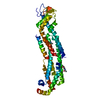 7zavC  7zawC  5fttS S: Starting model for refinement C: citing same article ( |
|---|---|
| Similar structure data |
-Links
 PDBj
PDBj


- Assembly
| Deposited unit | 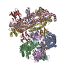
| ||||||||
|---|---|---|---|---|---|---|---|---|---|
| 1 |
| ||||||||
| Unit cell |
|
-Components
| #1: Protein | Mass: 53323.598 Da / Num. of mol.: 4 Source method: isolated from a genetically manipulated source Source: (gene. exp.)  Homo sapiens (human) / References: UniProt: Q8CFZ4 Homo sapiens (human) / References: UniProt: Q8CFZ4#2: Protein | Mass: 30257.984 Da / Num. of mol.: 4 Source method: isolated from a genetically manipulated source Source: (gene. exp.) Homo sapiens (human) / References: UniProt: F1LW30#3: Polysaccharide | 2-acetamido-2-deoxy-beta-D-glucopyranose-(1-4)-2-acetamido-2-deoxy-beta-D-glucopyranose | Source method: isolated from a genetically manipulated source #4: Sugar | ChemComp-NAG /   Type: D-saccharide, beta linking / Mass: 221.208 Da / Num. of mol.: 14 / Source method: obtained synthetically / Formula: C8H15NO6 Type: D-saccharide, beta linking / Mass: 221.208 Da / Num. of mol.: 14 / Source method: obtained synthetically / Formula: C8H15NO6#5: Sugar |   Type: D-saccharide, alpha linking / Mass: 180.156 Da / Num. of mol.: 3 / Source method: obtained synthetically / Formula: C6H12O6 Type: D-saccharide, alpha linking / Mass: 180.156 Da / Num. of mol.: 3 / Source method: obtained synthetically / Formula: C6H12O6Has ligand of interest | N | Has protein modification | Y | |
|---|
-Experimental details
-Experiment
| Experiment | Method: X-RAY DIFFRACTION / Number of used crystals: 1 |
|---|
- Sample preparation
| Crystal | Density Matthews: 3.18 Å3/Da / Density % sol: 61.38 % |
|---|---|
| Crystal grow | Temperature: 291.15 K / Method: vapor diffusion / pH: 7.8 Details: 0.1M Tris (pH 7.8) 5% w/v gamma-PGA, 20% w/vPEG 3350 |
-Data collection
| Diffraction | Mean temperature: 100 K / Serial crystal experiment: N |
|---|---|
| Diffraction source | Source: SYNCHROTRON / Site: Diamond / Beamline: I03 / Wavelength: 0.8 Å |
| Detector | Type: DECTRIS EIGER2 XE 16M / Detector: PIXEL / Date: Jul 9, 2017 |
| Radiation | Protocol: SINGLE WAVELENGTH / Monochromatic (M) / Laue (L): M / Scattering type: x-ray |
| Radiation wavelength | Wavelength: 0.8 Å / Relative weight: 1 |
| Reflection | Resolution: 4→80.878 Å / Num. obs: 26038 / % possible obs: 73.8 % / Redundancy: 8.2 % / Biso Wilson estimate: 269.6 Å2 / CC1/2: 1 / Rmerge(I) obs: 0.053 / Rpim(I) all: 0.02 / Net I/σ(I): 12.6 |
| Reflection shell | Resolution: 4→4.227 Å / Mean I/σ(I) obs: 2 / Num. unique obs: 1287 / CC1/2: 0.761 |
- Processing
| Software |
| |||||||||||||||||||||||||||||||||||||||||||||||||||||||||||||||||||||||||||||||||||||||||||||||||||||||||||||||||||||||||||||||||||||||||||||||||||||||||||||||||||||||||||||||||||||||||||||||||||||||||||||||||||||||||||||||||
|---|---|---|---|---|---|---|---|---|---|---|---|---|---|---|---|---|---|---|---|---|---|---|---|---|---|---|---|---|---|---|---|---|---|---|---|---|---|---|---|---|---|---|---|---|---|---|---|---|---|---|---|---|---|---|---|---|---|---|---|---|---|---|---|---|---|---|---|---|---|---|---|---|---|---|---|---|---|---|---|---|---|---|---|---|---|---|---|---|---|---|---|---|---|---|---|---|---|---|---|---|---|---|---|---|---|---|---|---|---|---|---|---|---|---|---|---|---|---|---|---|---|---|---|---|---|---|---|---|---|---|---|---|---|---|---|---|---|---|---|---|---|---|---|---|---|---|---|---|---|---|---|---|---|---|---|---|---|---|---|---|---|---|---|---|---|---|---|---|---|---|---|---|---|---|---|---|---|---|---|---|---|---|---|---|---|---|---|---|---|---|---|---|---|---|---|---|---|---|---|---|---|---|---|---|---|---|---|---|---|---|---|---|---|---|---|---|---|---|---|---|---|---|---|---|---|---|
| Refinement | Method to determine structure: MOLECULAR REPLACEMENT Starting model: 5FTT, GPC3 Resolution: 4→80.878 Å / Cor.coef. Fo:Fc: 0.861 / Cor.coef. Fo:Fc free: 0.875 / SU B: 457.993 / SU ML: 2.334 / Cross valid method: FREE R-VALUE / ESU R Free: 1.53 Details: Hydrogens have been added in their riding positions
| |||||||||||||||||||||||||||||||||||||||||||||||||||||||||||||||||||||||||||||||||||||||||||||||||||||||||||||||||||||||||||||||||||||||||||||||||||||||||||||||||||||||||||||||||||||||||||||||||||||||||||||||||||||||||||||||||
| Solvent computation | Ion probe radii: 0.8 Å / Shrinkage radii: 0.8 Å / VDW probe radii: 1.2 Å / Solvent model: MASK BULK SOLVENT | |||||||||||||||||||||||||||||||||||||||||||||||||||||||||||||||||||||||||||||||||||||||||||||||||||||||||||||||||||||||||||||||||||||||||||||||||||||||||||||||||||||||||||||||||||||||||||||||||||||||||||||||||||||||||||||||||
| Displacement parameters | Biso mean: 301.314 Å2
| |||||||||||||||||||||||||||||||||||||||||||||||||||||||||||||||||||||||||||||||||||||||||||||||||||||||||||||||||||||||||||||||||||||||||||||||||||||||||||||||||||||||||||||||||||||||||||||||||||||||||||||||||||||||||||||||||
| Refinement step | Cycle: LAST / Resolution: 4→80.878 Å
| |||||||||||||||||||||||||||||||||||||||||||||||||||||||||||||||||||||||||||||||||||||||||||||||||||||||||||||||||||||||||||||||||||||||||||||||||||||||||||||||||||||||||||||||||||||||||||||||||||||||||||||||||||||||||||||||||
| Refine LS restraints |
| |||||||||||||||||||||||||||||||||||||||||||||||||||||||||||||||||||||||||||||||||||||||||||||||||||||||||||||||||||||||||||||||||||||||||||||||||||||||||||||||||||||||||||||||||||||||||||||||||||||||||||||||||||||||||||||||||
| LS refinement shell |
| |||||||||||||||||||||||||||||||||||||||||||||||||||||||||||||||||||||||||||||||||||||||||||||||||||||||||||||||||||||||||||||||||||||||||||||||||||||||||||||||||||||||||||||||||||||||||||||||||||||||||||||||||||||||||||||||||
| Refinement TLS params. | Method: refined / Refine-ID: X-RAY DIFFRACTION
| |||||||||||||||||||||||||||||||||||||||||||||||||||||||||||||||||||||||||||||||||||||||||||||||||||||||||||||||||||||||||||||||||||||||||||||||||||||||||||||||||||||||||||||||||||||||||||||||||||||||||||||||||||||||||||||||||
| Refinement TLS group |
|
