 Movie
Movie Controller

[English] 日本語
 Yorodumi
- PDB-7z8o: Crystal structure of SARS-CoV-2 S RBD in complex with a stapled p... -
+ Open data

- Basic information
| Entry | Database: PDB / ID: 7z8o | ||||||
|---|---|---|---|---|---|---|---|
| Title | Crystal structure of SARS-CoV-2 S RBD in complex with a stapled peptide | ||||||
 Components Components |
| ||||||
 Keywords Keywords | VIRAL PROTEIN / sRBD / Stapled peptide | ||||||
| Function / homology |  Function and homology information Function and homology informationsymbiont-mediated disruption of host tissue / Maturation of spike protein / Translation of Structural Proteins / Virion Assembly and Release / host cell surface / host extracellular region / symbiont-mediated-mediated suppression of host tetherin activity / Induction of Cell-Cell Fusion / structural constituent of virion / positive regulation of viral entry into host cell ...symbiont-mediated disruption of host tissue / Maturation of spike protein / Translation of Structural Proteins / Virion Assembly and Release / host cell surface / host extracellular region / symbiont-mediated-mediated suppression of host tetherin activity / Induction of Cell-Cell Fusion / structural constituent of virion / positive regulation of viral entry into host cell / membrane fusion / host cell endoplasmic reticulum-Golgi intermediate compartment membrane / Attachment and Entry / entry receptor-mediated virion attachment to host cell / receptor-mediated virion attachment to host cell / host cell surface receptor binding / symbiont-mediated suppression of host innate immune response / endocytosis involved in viral entry into host cell / receptor ligand activity / fusion of virus membrane with host plasma membrane / fusion of virus membrane with host endosome membrane / viral envelope / symbiont entry into host cell / virion attachment to host cell / host cell plasma membrane / SARS-CoV-2 activates/modulates innate and adaptive immune responses / virion membrane / membrane / identical protein binding / plasma membrane Similarity search - Function | ||||||
| Biological species |   Severe acute respiratory syndrome coronavirus 2 Severe acute respiratory syndrome coronavirus 2synthetic construct (others) | ||||||
| Method |  X-RAY DIFFRACTION / SYNCHROTRON / MOLECULAR REPLACEMENT / Resolution: 0.96 Å X-RAY DIFFRACTION / SYNCHROTRON / MOLECULAR REPLACEMENT / Resolution: 0.96 Å | ||||||
 Authors Authors | Brear, P. / Chen, L. / Gaynor, K. / Harman, M. / Dods, R. / Hyvonen, M. | ||||||
| Funding support | 1items
| ||||||
 Citation Citation | Journal: Nat Commun / Year: 2023 Title: Multivalent bicyclic peptides are an effective antiviral modality that can potently inhibit SARS-CoV-2. Authors: Gaynor, K.U. / Vaysburd, M. / Harman, M.A.J. / Albecka, A. / Jeffrey, P. / Beswick, P. / Papa, G. / Chen, L. / Mallery, D. / McGuinness, B. / Van Rietschoten, K. / Stanway, S. / Brear, P. / ...Authors: Gaynor, K.U. / Vaysburd, M. / Harman, M.A.J. / Albecka, A. / Jeffrey, P. / Beswick, P. / Papa, G. / Chen, L. / Mallery, D. / McGuinness, B. / Van Rietschoten, K. / Stanway, S. / Brear, P. / Lulla, A. / Ciazynska, K. / Chang, V.T. / Sharp, J. / Neary, M. / Box, H. / Herriott, J. / Kijak, E. / Tatham, L. / Bentley, E.G. / Sharma, P. / Kirby, A. / Han, X. / Stewart, J.P. / Owen, A. / Briggs, J.A.G. / Hyvonen, M. / Skynner, M.J. / James, L.C. | ||||||
| History |
|
- Structure visualization
| Structure viewer | Molecule: MolmilJmol/JSmol |
|---|
- Downloads & links
-Download
| PDBx/mmCIF format | 7z8o.cif.gz | 120.4 KB | Display | PDBx/mmCIF format |
|---|---|---|---|---|
| PDB format | pdb7z8o.ent.gz | 90.6 KB | Display | PDB format |
| PDBx/mmJSON format | 7z8o.json.gz | Tree view | PDBx/mmJSON format | |
| Others |  Other downloads Other downloads |
-Validation report
| Arichive directory | https://data.pdbj.org/pub/pdb/validation_reports/z8/7z8oftp://data.pdbj.org/pub/pdb/validation_reports/z8/7z8o | HTTPS FTP |
|---|
-Related structure data
| Related structure data | 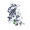 8aaaC 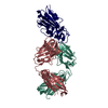 7ch5S S: Starting model for refinement C: citing same article ( |
|---|---|
| Similar structure data |
-Links
 PDBj
PDBj


- Assembly
| Deposited unit | 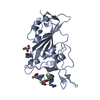
| ||||||||
|---|---|---|---|---|---|---|---|---|---|
| 1 |
| ||||||||
| Unit cell |
|
-Components
| #1: Protein | Mass: 22074.678 Da / Num. of mol.: 1 Source method: isolated from a genetically manipulated source Source: (gene. exp.) Severe acute respiratory syndrome coronavirus 2Gene: S, 2 / Production host:  | ||||||
|---|---|---|---|---|---|---|---|
| #2: Protein/peptide | Mass: 1395.736 Da / Num. of mol.: 1 / Source method: obtained synthetically / Source: (synth.) synthetic construct (others) | ||||||
| #3: Chemical |   Mass: 92.094 Da / Num. of mol.: 3 / Source method: obtained synthetically / Formula: C3H8O3 / Feature type: SUBJECT OF INVESTIGATION Mass: 92.094 Da / Num. of mol.: 3 / Source method: obtained synthetically / Formula: C3H8O3 / Feature type: SUBJECT OF INVESTIGATION#4: Chemical | ChemComp-KZ0 / |   Mass: 226.491 Da / Num. of mol.: 1 / Source method: obtained synthetically / Formula: C6H6Cl3N3 / Feature type: SUBJECT OF INVESTIGATION Mass: 226.491 Da / Num. of mol.: 1 / Source method: obtained synthetically / Formula: C6H6Cl3N3 / Feature type: SUBJECT OF INVESTIGATION#5: Water | ChemComp-HOH / |  Mass: 18.015 Da / Num. of mol.: 340 / Source method: isolated from a natural source / Formula: H2O Mass: 18.015 Da / Num. of mol.: 340 / Source method: isolated from a natural source / Formula: H2OHas ligand of interest | Y | |
-Experimental details
-Experiment
| Experiment | Method: X-RAY DIFFRACTION / Number of used crystals: 1 |
|---|
- Sample preparation
| Crystal | Density Matthews: 2.23 Å3/Da / Density % sol: 44.92 % |
|---|---|
| Crystal grow | Temperature: 292 K / Method: vapor diffusion, sitting drop / Details: 22 %v/v PEGSB, 0.1 M Na Phos Cit 5.5 pH |
-Data collection
| Diffraction | Mean temperature: 100 K / Serial crystal experiment: N | ||||||||||||||||||||||||||||||
|---|---|---|---|---|---|---|---|---|---|---|---|---|---|---|---|---|---|---|---|---|---|---|---|---|---|---|---|---|---|---|---|
| Diffraction source | Source: SYNCHROTRON / Site: Diamond  / Beamline: I04 / Wavelength: 0.8 Å / Beamline: I04 / Wavelength: 0.8 Å | ||||||||||||||||||||||||||||||
| Detector | Type: DECTRIS EIGER2 XE 16M / Detector: PIXEL / Date: Feb 24, 2021 | ||||||||||||||||||||||||||||||
| Radiation | Protocol: SINGLE WAVELENGTH / Monochromatic (M) / Laue (L): M / Scattering type: x-ray | ||||||||||||||||||||||||||||||
| Radiation wavelength | Wavelength: 0.8 Å / Relative weight: 1 | ||||||||||||||||||||||||||||||
| Reflection | Resolution: 0.96→46.2 Å / Num. obs: 123986 / % possible obs: 99.2 % / Redundancy: 18.7 % / CC1/2: 1 / Rmerge(I) obs: 0.061 / Rpim(I) all: 0.014 / Rrim(I) all: 0.062 / Net I/σ(I): 19.6 / Num. measured all: 2313853 / Scaling rejects: 408 | ||||||||||||||||||||||||||||||
| Reflection shell | Diffraction-ID: 1
|
- Processing
| Software |
| |||||||||||||||||||||||||||||||||||||||||||||||||||||||||||||||||
|---|---|---|---|---|---|---|---|---|---|---|---|---|---|---|---|---|---|---|---|---|---|---|---|---|---|---|---|---|---|---|---|---|---|---|---|---|---|---|---|---|---|---|---|---|---|---|---|---|---|---|---|---|---|---|---|---|---|---|---|---|---|---|---|---|---|---|
| Refinement | Method to determine structure: MOLECULAR REPLACEMENT Starting model: 7CH5 Resolution: 0.96→46.2 Å / Cor.coef. Fo:Fc: 0.98 / Cor.coef. Fo:Fc free: 0.978 / SU B: 0.798 / SU ML: 0.019 / Cross valid method: THROUGHOUT / σ(F): 0 / ESU R: 0.022 / ESU R Free: 0.023 / Stereochemistry target values: MAXIMUM LIKELIHOOD Details: HYDROGENS HAVE BEEN ADDED IN THE RIDING POSITIONS U VALUES : REFINED INDIVIDUALLY
| |||||||||||||||||||||||||||||||||||||||||||||||||||||||||||||||||
| Solvent computation | Ion probe radii: 0.8 Å / Shrinkage radii: 0.8 Å / VDW probe radii: 1.2 Å / Solvent model: MASK | |||||||||||||||||||||||||||||||||||||||||||||||||||||||||||||||||
| Displacement parameters | Biso max: 115.42 Å2 / Biso mean: 15.855 Å2 / Biso min: 6.71 Å2
| |||||||||||||||||||||||||||||||||||||||||||||||||||||||||||||||||
| Refinement step | Cycle: final / Resolution: 0.96→46.2 Å
| |||||||||||||||||||||||||||||||||||||||||||||||||||||||||||||||||
| Refine LS restraints |
| |||||||||||||||||||||||||||||||||||||||||||||||||||||||||||||||||
| LS refinement shell | Resolution: 0.96→0.985 Å / Rfactor Rfree error: 0 / Total num. of bins used: 20
|
