[English] 日本語
Yorodumi
- PDB-7z8n: GacS histidine kinase from Pseudomonas aeruginosa -

+
Open data


ID or keywords:

Loading...

-
Basic information

Entry
Database: PDB / ID: 7z8n
TitleGacS histidine kinase from Pseudomonas aeruginosa
ComponentsHistidine kinase
KeywordsSIGNALING PROTEIN / Histidine Kinase HAMP domain Coiled-coil GacS Pseudomonas aeruginosa
Function / homology
Function and homology information


positive regulation of secondary metabolite biosynthetic process / positive regulation of cell motility / histidine kinase / phosphorelay sensor kinase activity / plasma membrane
Similarity search - Function
Histidine kinase BarA, N-terminal / Single cache domain 4 / Histidine Phosphotransfer domain / Hpt domain / Histidine-containing phosphotransfer (HPt) domain profile. / Signal transduction histidine kinase, phosphotransfer (Hpt) domain / HPT domain superfamily / HAMP domain / HAMP (Histidine kinases, Adenylyl cyclases, Methyl binding proteins, Phosphatases) domain / HAMP domain profile. ...Histidine kinase BarA, N-terminal / Single cache domain 4 / Histidine Phosphotransfer domain / Hpt domain / Histidine-containing phosphotransfer (HPt) domain profile. / Signal transduction histidine kinase, phosphotransfer (Hpt) domain / HPT domain superfamily / HAMP domain / HAMP (Histidine kinases, Adenylyl cyclases, Methyl binding proteins, Phosphatases) domain / HAMP domain profile. / HAMP domain / His Kinase A (phospho-acceptor) domain / His Kinase A (phosphoacceptor) domain / Signal transduction histidine kinase, dimerisation/phosphoacceptor domain / Signal transduction histidine kinase, dimerisation/phosphoacceptor domain superfamily / Signal transduction histidine kinase-related protein, C-terminal / Histidine kinase domain / Histidine kinase domain profile. / Response regulator receiver domain / cheY-homologous receiver domain / Signal transduction response regulator, receiver domain / Response regulatory domain profile. / CheY-like superfamily / Histidine kinase-, DNA gyrase B-, and HSP90-like ATPase / Histidine kinase-like ATPases / Histidine kinase/HSP90-like ATPase / Histidine kinase/HSP90-like ATPase superfamily
Similarity search - Domain/homology
R-1,2-PROPANEDIOL / histidine kinase
Similarity search - Component
Biological speciesPseudomonas aeruginosa PAO1 (bacteria)
MethodX-RAY DIFFRACTION / SYNCHROTRON / SAD / Resolution: 2.64 Å
AuthorsFadel, F. / Bassim, V. / Francis, V.I. / Porter, S.L. / Botzanowski, T. / Legrand, P. / Bourne, Y. / Cianferani, S. / Vincent, F.
Funding support France, 1items
OrganizationGrant numberCountry
Agence Nationale de la Recherche (ANR) France
CitationJournal: Structure / Year: 2022
Title: Insights into the atypical autokinase activity of the Pseudomonas aeruginosa GacS histidine kinase and its interaction with RetS.
Authors: Fadel, F. / Bassim, V. / Francis, V.I. / Porter, S.L. / Botzanowski, T. / Legrand, P. / Perez, M.M. / Bourne, Y. / Cianferani, S. / Vincent, F.
History
DepositionMar 17, 2022Deposition site: PDBE / Processing site: PDBE
SupersessionJul 13, 2022ID: 6ZLS
Revision 1.0Jul 13, 2022Provider: repository / Type: Initial release
Revision 1.1Jan 25, 2023Group: Database references / Category: citation / citation_author
Item: _citation.country / _citation.journal_abbrev ..._citation.country / _citation.journal_abbrev / _citation.journal_id_ASTM / _citation.journal_id_CSD / _citation.journal_id_ISSN / _citation.journal_volume / _citation.page_first / _citation.page_last / _citation.pdbx_database_id_DOI / _citation.pdbx_database_id_PubMed / _citation.title / _citation.year

-
Structure visualization

Structure viewerMolecule:
MolmilJmol/JSmol

Downloads & links

-
Assembly

Deposited unit
A: Histidine kinase
B: Histidine kinase
C: Histidine kinase
D: Histidine kinase
hetero molecules


Theoretical massNumber of molelcules
Total (without water)141,90610
Polymers141,5584
Non-polymers3496
Water3,207178
1


  • Idetical with deposited unit
  • defined by author
  • Evidence: mass spectrometry
TypeNameSymmetry operationNumber
identity operation1_555x,y,z1
Unit cell
Length a, b, c (Å)63.710, 76.850, 178.650
Angle α, β, γ (deg.)90.000, 97.300, 90.000
Int Tables number4
Space group name H-MP1211

-
Components

#1: Protein
Histidine kinase


Mass: 35389.383 Da / Num. of mol.: 4
Source method: isolated from a genetically manipulated source
Details: The protein sequence has been produced with a histidine tag followed by a TEV protease cleavage site. This part of the protein is not modelled and does not appear in the PDB sequence.
Source: (gene. exp.) Pseudomonas aeruginosa PAO1 (bacteria)
Strain: ATCC 15692 / DSM 22644 / CIP 104116 / JCM 14847 / LMG 12228 / 1C / PRS 101 / PAO1
Gene: gacS, PA0928 / Plasmid: pLIC03 / Production host: Escherichia coli BL21(DE3) (bacteria) / References: UniProt: G3XD98, histidine kinase
#2: Chemical ChemComp-CA / CALCIUM ION


Mass: 40.078 Da / Num. of mol.: 3 / Source method: obtained synthetically / Formula: Ca
#3: Chemical ChemComp-PGR / R-1,2-PROPANEDIOL


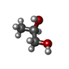
Mass: 76.094 Da / Num. of mol.: 3 / Source method: obtained synthetically / Formula: C3H8O2
#4: Water ChemComp-HOH / water


Mass: 18.015 Da / Num. of mol.: 178 / Source method: isolated from a natural source / Formula: H2O
Has ligand of interestN

-
Experimental details

-
Experiment

ExperimentMethod: X-RAY DIFFRACTION / Number of used crystals: 1

-
Sample preparation

CrystalDensity Matthews: 3.06 Å3/Da / Density % sol: 59.86 %
Crystal growTemperature: 293 K / Method: vapor diffusion, sitting drop / Details: 0.2M Lithium chloride 24% PEG 3350 0.1M MES pH 5.5

-
Data collection

DiffractionMean temperature: 100 K / Serial crystal experiment: N
Diffraction sourceSource: SYNCHROTRON / Site: SOLEIL / Beamline: PROXIMA 1 / Wavelength: 0.9794 Å
DetectorType: DECTRIS EIGER X 16M / Detector: PIXEL / Date: Jul 11, 2017
RadiationProtocol: MAD / Monochromatic (M) / Laue (L): M / Scattering type: x-ray
Radiation wavelengthWavelength: 0.9794 Å / Relative weight: 1
ReflectionResolution: 2.64→48.81 Å / Num. obs: 45042 / % possible obs: 89 % / Redundancy: 13.6 % / CC1/2: 0.998 / Net I/σ(I): 16
Reflection shellResolution: 2.64→2.71 Å / Num. unique obs: 2252 / CC1/2: 0.429

-
Processing

Software
NameVersionClassification
BUSTER2.10.4 (3-FEB-2022)refinement
PDB_EXTRACT3.27data extraction
XDSdata reduction
Aimlessdata scaling
SHELXCDphasing
RefinementMethod to determine structure: SAD / Resolution: 2.64→48.81 Å / Cor.coef. Fo:Fc: 0.922 / Cor.coef. Fo:Fc free: 0.933 / SU R Cruickshank DPI: 0.773 / Cross valid method: THROUGHOUT / σ(F): 0 / SU R Blow DPI: 0.705 / SU Rfree Blow DPI: 0.318 / SU Rfree Cruickshank DPI: 0.329
RfactorNum. reflection% reflectionSelection details
Rfree0.2641 2266 5.03 %RANDOM
Rwork0.2592 ---
obs0.2594 45042 89 %-
Displacement parametersBiso max: 257.25 Å2 / Biso mean: 95.9 Å2 / Biso min: 35.79 Å2
Baniso -1Baniso -2Baniso -3
1-3.1734 Å20 Å21.0352 Å2
2---5.7291 Å20 Å2
3---2.5557 Å2
Refine analyzeLuzzati coordinate error obs: 0.45 Å
Refinement stepCycle: final / Resolution: 2.64→48.81 Å
ProteinNucleic acidLigandSolventTotal
Num. atoms9032 0 18 178 9228
Biso mean--91.47 67.18 -
Num. residues----1168
Refine LS restraints
Refine-IDTypeNumberRestraint functionWeightDev ideal
X-RAY DIFFRACTIONt_dihedral_angle_d3447SINUSOIDAL2
X-RAY DIFFRACTIONt_trig_c_planes
X-RAY DIFFRACTIONt_gen_planes1639HARMONIC5
X-RAY DIFFRACTIONt_it9213HARMONIC10
X-RAY DIFFRACTIONt_nbd
X-RAY DIFFRACTIONt_improper_torsion
X-RAY DIFFRACTIONt_pseud_angle
X-RAY DIFFRACTIONt_chiral_improper_torsion1254SEMIHARMONIC5
X-RAY DIFFRACTIONt_sum_occupancies
X-RAY DIFFRACTIONt_utility_distance
X-RAY DIFFRACTIONt_utility_angle
X-RAY DIFFRACTIONt_utility_torsion
X-RAY DIFFRACTIONt_ideal_dist_contact6912SEMIHARMONIC4
X-RAY DIFFRACTIONt_bond_d9213HARMONIC20.007
X-RAY DIFFRACTIONt_angle_deg12447HARMONIC20.87
X-RAY DIFFRACTIONt_omega_torsion2.45
X-RAY DIFFRACTIONt_other_torsion19.15
LS refinement shellResolution: 2.64→2.67 Å / Rfactor Rfree error: 0 / Total num. of bins used: 51
RfactorNum. reflection% reflection
Rfree0.4221 45 4.99 %
Rwork0.3253 856 -
all0.33 901 -
obs--46.41 %
Refinement TLS params.

Method: refined / Refine-ID: X-RAY DIFFRACTION

IDL112)L122)L132)L222)L232)L332)S11 (Å °)S12 (Å °)S13 (Å °)S21 (Å °)S22 (Å °)S23 (Å °)S31 (Å °)S32 (Å °)S33 (Å °)T112)T122)T132)T222)T232)T332)Origin x (Å)Origin y (Å)Origin z (Å)
10.4098-0.36840.29790.91340.6310.9205-0.00850.31120.0564-0.1124-0.0879-0.0118-0.11750.21530.09640.0030.10340.05030.03130.1197-0.0778-110.835335.0503144.1506
21.12230.11980.01310.2714-0.08270.09230.0826-0.4015-0.0748-0.0501-0.03230.0333-0.00770.0681-0.0502-0.013-0.03920.05680.01420.0055-0.0179-7.953836.057327.2031
Refinement TLS group
IDRefine-IDRefine TLS-IDSelection detailsAuth asym-IDAuth seq-ID
1X-RAY DIFFRACTION1{ A|256 - A|512 D|256 - D|512 }A256 - 512
2X-RAY DIFFRACTION1{ A|256 - A|512 D|256 - D|512 }D256 - 512
3X-RAY DIFFRACTION2{ B|256 - B|512 C|256 - C|512 }B256 - 512
4X-RAY DIFFRACTION2{ B|256 - B|512 C|256 - C|512 }C256 - 512

+
About Yorodumi

-
News

-
Feb 9, 2022. New format data for meta-information of EMDB entries

New format data for meta-information of EMDB entries

  • Version 3 of the EMDB header file is now the official format.
  • The previous official version 1.9 will be removed from the archive.

Related info.:EMDB header

External links:wwPDB to switch to version 3 of the EMDB data model

-
Aug 12, 2020. Covid-19 info

Covid-19 info

URL: https://pdbjlvh1.pdbj.org/emnavi/covid19.php

New page: Covid-19 featured information page in EM Navigator.

Related info.:Covid-19 info / Mar 5, 2020. Novel coronavirus structure data

+
Mar 5, 2020. Novel coronavirus structure data

Novel coronavirus structure data

Related info.:Yorodumi Speices / Aug 12, 2020. Covid-19 info

External links:COVID-19 featured content - PDBj / Molecule of the Month (242):Coronavirus Proteases

+
Jan 31, 2019. EMDB accession codes are about to change! (news from PDBe EMDB page)

EMDB accession codes are about to change! (news from PDBe EMDB page)

  • The allocation of 4 digits for EMDB accession codes will soon come to an end. Whilst these codes will remain in use, new EMDB accession codes will include an additional digit and will expand incrementally as the available range of codes is exhausted. The current 4-digit format prefixed with “EMD-” (i.e. EMD-XXXX) will advance to a 5-digit format (i.e. EMD-XXXXX), and so on. It is currently estimated that the 4-digit codes will be depleted around Spring 2019, at which point the 5-digit format will come into force.
  • The EM Navigator/Yorodumi systems omit the EMD- prefix.

Related info.:Q: What is EMD? / ID/Accession-code notation in Yorodumi/EM Navigator

External links:EMDB Accession Codes are Changing Soon! / Contact to PDBj

+
Jul 12, 2017. Major update of PDB

Major update of PDB

  • wwPDB released updated PDB data conforming to the new PDBx/mmCIF dictionary.
  • This is a major update changing the version number from 4 to 5, and with Remediation, in which all the entries are updated.
  • In this update, many items about electron microscopy experimental information are reorganized (e.g. em_software).
  • Now, EM Navigator and Yorodumi are based on the updated data.

External links:wwPDB Remediation / Enriched Model Files Conforming to OneDep Data Standards Now Available in the PDB FTP Archive

-
Yorodumi

Thousand views of thousand structures

  • Yorodumi is a browser for structure data from EMDB, PDB, SASBDB, etc.
  • This page is also the successor to EM Navigator detail page, and also detail information page/front-end page for Omokage search.
  • The word "yorodu" (or yorozu) is an old Japanese word meaning "ten thousand". "mi" (miru) is to see.

Related info.:EMDB / PDB / SASBDB / Comparison of 3 databanks / Yorodumi Search / Aug 31, 2016. New EM Navigator & Yorodumi / Yorodumi Papers / Jmol/JSmol / Function and homology information / Changes in new EM Navigator and Yorodumi

Read more