Movie
Movie Controller


+ Open data

- Basic information
| Entry | Database: PDB / ID: 7z7h | |||||||||
|---|---|---|---|---|---|---|---|---|---|---|
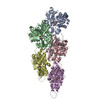
| Title | Structure of P. luminescens TccC3-F-actin complex | |||||||||
 Components Components |
| |||||||||
 Keywords Keywords | TOXIN / Bacterial toxin / F-actin | |||||||||
| Function / homology |  Function and homology information Function and homology informationcytoskeletal motor activator activity / myosin heavy chain binding / tropomyosin binding / actin filament bundle / detection of maltose stimulus / troponin I binding / filamentous actin / maltose transport complex / mesenchyme migration / carbohydrate transport ...cytoskeletal motor activator activity / myosin heavy chain binding / tropomyosin binding / actin filament bundle / detection of maltose stimulus / troponin I binding / filamentous actin / maltose transport complex / mesenchyme migration / carbohydrate transport / skeletal muscle myofibril / actin filament bundle assembly / striated muscle thin filament / skeletal muscle thin filament assembly / actin monomer binding / carbohydrate transmembrane transporter activity / maltose binding / maltose transport / maltodextrin transmembrane transport / ATP-binding cassette (ABC) transporter complex, substrate-binding subunit-containing / skeletal muscle fiber development / stress fiber / titin binding / actin filament polymerization / ATP-binding cassette (ABC) transporter complex / cell chemotaxis / filopodium / actin filament / Hydrolases; Acting on acid anhydrides; Acting on acid anhydrides to facilitate cellular and subcellular movement / calcium-dependent protein binding / lamellipodium / outer membrane-bounded periplasmic space / cell body / periplasmic space / protein domain specific binding / hydrolase activity / calcium ion binding / DNA damage response / positive regulation of gene expression / magnesium ion binding / ATP binding / membrane / identical protein binding / cytoplasm Similarity search - Function | |||||||||
| Biological species |  Photorhabdus luminescens (bacteria) Photorhabdus luminescens (bacteria) | |||||||||
| Method | ELECTRON MICROSCOPY / single particle reconstruction / cryo EM / Resolution: 3.8 Å | |||||||||
 Authors Authors | Belyy, A. / Raunser, S. | |||||||||
| Funding support |  Germany, 1items Germany, 1items
| |||||||||
 Citation Citation | Journal: Nat Commun / Year: 2022 Title: Mechanism of threonine ADP-ribosylation of F-actin by a Tc toxin. Authors: Alexander Belyy / Florian Lindemann / Daniel Roderer / Johanna Funk / Benjamin Bardiaux / Jonas Protze / Peter Bieling / Hartmut Oschkinat / Stefan Raunser /  Abstract: Tc toxins deliver toxic enzymes into host cells by a unique injection mechanism. One of these enzymes is the actin ADP-ribosyltransferase TccC3, whose activity leads to the clustering of the cellular ...Tc toxins deliver toxic enzymes into host cells by a unique injection mechanism. One of these enzymes is the actin ADP-ribosyltransferase TccC3, whose activity leads to the clustering of the cellular cytoskeleton and ultimately cell death. Here, we show in atomic detail how TccC3 modifies actin. We find that the ADP-ribosyltransferase does not bind to G-actin but interacts with two consecutive actin subunits of F-actin. The binding of TccC3 to F-actin occurs via an induced-fit mechanism that facilitates access of NAD to the nucleotide binding pocket. The following nucleophilic substitution reaction results in the transfer of ADP-ribose to threonine-148 of F-actin. We demonstrate that this site-specific modification of F-actin prevents its interaction with depolymerization factors, such as cofilin, which impairs actin network turnover and leads to steady actin polymerization. Our findings reveal in atomic detail a mechanism of action of a bacterial toxin through specific targeting and modification of F-actin. | |||||||||
| History |
|
- Structure visualization
| Structure viewer | Molecule: MolmilJmol/JSmol |
|---|
- Downloads & links
-Download
| PDBx/mmCIF format | 7z7h.cif.gz | 364 KB | Display | PDBx/mmCIF format |
|---|---|---|---|---|
| PDB format | pdb7z7h.ent.gz | 296.3 KB | Display | PDB format |
| PDBx/mmJSON format | 7z7h.json.gz | Tree view | PDBx/mmJSON format | |
| Others |  Other downloads Other downloads |
-Validation report
| Arichive directory | https://data.pdbj.org/pub/pdb/validation_reports/z7/7z7hftp://data.pdbj.org/pub/pdb/validation_reports/z7/7z7h | HTTPS FTP |
|---|
-Related structure data
| Related structure data | 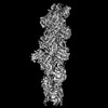 14532MC  7z7iC 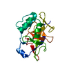 7zbqC M: map data used to model this data C: citing same article ( |
|---|---|
| Similar structure data |

-Links
 PDBj
PDBj


- Assembly
| Deposited unit | 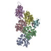
|
|---|---|
| 1 |
|
-Components
-Protein , 2 types, 6 molecules DBECAF
| #1: Protein | Mass: 42109.973 Da / Num. of mol.: 5 / Source method: isolated from a natural source / Source: (natural) #2: Protein | | Mass: 63658.949 Da / Num. of mol.: 1 Source method: isolated from a genetically manipulated source Source: (gene. exp.) Photorhabdus luminescens (bacteria) / Strain: K12 / Gene: malE, b4034, JW3994, TccC3 / Production host: |
|---|
-Non-polymers , 4 types, 12 molecules 


| #3: Chemical | ChemComp-ADP /  Mass: 427.201 Da / Num. of mol.: 5 / Source method: obtained synthetically / Formula: C10H15N5O10P2 / Comment: ADP, energy-carrying molecule*YM Mass: 427.201 Da / Num. of mol.: 5 / Source method: obtained synthetically / Formula: C10H15N5O10P2 / Comment: ADP, energy-carrying molecule*YM#4: Chemical | ChemComp-MG /  Mass: 24.305 Da / Num. of mol.: 5 / Source method: obtained synthetically / Formula: Mg Mass: 24.305 Da / Num. of mol.: 5 / Source method: obtained synthetically / Formula: Mg#5: Chemical | ChemComp-APR / |  Mass: 559.316 Da / Num. of mol.: 1 / Source method: obtained synthetically / Formula: C15H23N5O14P2 / Feature type: SUBJECT OF INVESTIGATION Mass: 559.316 Da / Num. of mol.: 1 / Source method: obtained synthetically / Formula: C15H23N5O14P2 / Feature type: SUBJECT OF INVESTIGATION#6: Chemical | ChemComp-NCA / |  Mass: 122.125 Da / Num. of mol.: 1 / Source method: obtained synthetically / Formula: C6H6N2O / Feature type: SUBJECT OF INVESTIGATION / Comment: medication*YM Mass: 122.125 Da / Num. of mol.: 1 / Source method: obtained synthetically / Formula: C6H6N2O / Feature type: SUBJECT OF INVESTIGATION / Comment: medication*YM |
|---|
-Details
| Has ligand of interest | Y |
|---|
-Experimental details
-Experiment
| Experiment | Method: ELECTRON MICROSCOPY |
|---|---|
| EM experiment | Aggregation state: FILAMENT / 3D reconstruction method: single particle reconstruction |
- Sample preparation
| Component | Name: Structure of P. luminescence TccC3-F-actin complex / Type: COMPLEX / Entity ID: #1-#2 / Source: RECOMBINANT |
|---|---|
| Molecular weight | Experimental value: NO |
| Source (natural) | Organism: Photorhabdus luminescens (bacteria) |
| Source (recombinant) | Organism: |
| Buffer solution | pH: 8 |
| Specimen | Embedding applied: NO / Shadowing applied: NO / Staining applied: NO / Vitrification applied: YES |
| Specimen support | Grid material: COPPER / Grid mesh size: 300 divisions/in. / Grid type: Quantifoil R2/1 |
| Vitrification | Instrument: FEI VITROBOT MARK III / Cryogen name: ETHANE / Humidity: 100 % |
- Electron microscopy imaging
| Experimental equipment |  Model: Titan Krios / Image courtesy: FEI Company |
|---|---|
| Microscopy | Model: FEI TITAN KRIOS |
| Electron gun | Electron source:  FIELD EMISSION GUN / Accelerating voltage: 300 kV / Illumination mode: SPOT SCAN FIELD EMISSION GUN / Accelerating voltage: 300 kV / Illumination mode: SPOT SCAN |
| Electron lens | Mode: BRIGHT FIELD / Nominal defocus max: 2800 nm / Nominal defocus min: 400 nm |
| Image recording | Electron dose: 84.9 e/Å2 / Film or detector model: GATAN K3 (6k x 4k) / Num. of grids imaged: 1 / Num. of real images: 12284 |
- Processing
| Software | Name: PHENIX / Version: 1.17.1_3660: / Classification: refinement | ||||||||||||||||||||||||||||||||||||
|---|---|---|---|---|---|---|---|---|---|---|---|---|---|---|---|---|---|---|---|---|---|---|---|---|---|---|---|---|---|---|---|---|---|---|---|---|---|
| EM software |
| ||||||||||||||||||||||||||||||||||||
| CTF correction | Type: PHASE FLIPPING AND AMPLITUDE CORRECTION | ||||||||||||||||||||||||||||||||||||
| Particle selection | Num. of particles selected: 2224805 | ||||||||||||||||||||||||||||||||||||
| Symmetry | Point symmetry: C1 (asymmetric) | ||||||||||||||||||||||||||||||||||||
| 3D reconstruction | Resolution: 3.8 Å / Resolution method: FSC 0.143 CUT-OFF / Num. of particles: 437658 / Algorithm: FOURIER SPACE / Symmetry type: POINT | ||||||||||||||||||||||||||||||||||||
| Atomic model building | Protocol: FLEXIBLE FIT / Space: REAL | ||||||||||||||||||||||||||||||||||||
| Atomic model building | PDB-ID: 5ONV | ||||||||||||||||||||||||||||||||||||
| Refine LS restraints |
|