 Movie
Movie Controller

[English] 日本語
 Yorodumi
- PDB-7z5a: Crystal structure of the trapped complex of mouse Endonuclease VI... -
+ Open data

- Basic information
| Entry | Database: PDB / ID: 7z5a | ||||||
|---|---|---|---|---|---|---|---|
| Title | Crystal structure of the trapped complex of mouse Endonuclease VIII-LIKE 3 (mNEIL3) and hairpin DNA with 5'overhang | ||||||
 Components Components |
| ||||||
 Keywords Keywords | DNA BINDING PROTEIN / Ap-ICL / DNA crosslink repair / NEIL3 / BER | ||||||
| Function / homology |  Function and homology information Function and homology informationRecognition and association of DNA glycosylase with site containing an affected purine / Cleavage of the damaged purine / MCM complex binding / DNA N-glycosylase activity / single strand break repair / bubble DNA binding / Hydrolases; Glycosylases; Hydrolysing N-glycosyl compounds / interstrand cross-link repair / DNA-(apurinic or apyrimidinic site) endonuclease activity / DNA-(apurinic or apyrimidinic site) lyase ...Recognition and association of DNA glycosylase with site containing an affected purine / Cleavage of the damaged purine / MCM complex binding / DNA N-glycosylase activity / single strand break repair / bubble DNA binding / Hydrolases; Glycosylases; Hydrolysing N-glycosyl compounds / interstrand cross-link repair / DNA-(apurinic or apyrimidinic site) endonuclease activity / DNA-(apurinic or apyrimidinic site) lyase / class I DNA-(apurinic or apyrimidinic site) endonuclease activity / base-excision repair / single-stranded DNA binding / chromosome / double-stranded DNA binding / damaged DNA binding / zinc ion binding / nucleoplasm / nucleus Similarity search - Function | ||||||
| Biological species |  synthetic construct (others) | ||||||
| Method |  X-RAY DIFFRACTION / SYNCHROTRON / MOLECULAR REPLACEMENT / Resolution: 2.28 Å X-RAY DIFFRACTION / SYNCHROTRON / MOLECULAR REPLACEMENT / Resolution: 2.28 Å | ||||||
 Authors Authors | Silhan, J. / Huskova, A. | ||||||
| Funding support |  Czech Republic, 1items Czech Republic, 1items
| ||||||
 Citation Citation | Journal: Nucleic Acids Res. / Year: 2022 Title: Model of abasic site DNA cross-link repair; from the architecture of NEIL3 DNA binding domains to the X-structure model. Authors: Huskova, A. / Dinesh, D.C. / Srb, P. / Boura, E. / Veverka, V. / Silhan, J. | ||||||
| History |
|
- Structure visualization
| Structure viewer | Molecule: MolmilJmol/JSmol |
|---|
- Downloads & links
-Download
| PDBx/mmCIF format | 7z5a.cif.gz | 81.1 KB | Display | PDBx/mmCIF format |
|---|---|---|---|---|
| PDB format | pdb7z5a.ent.gz | 55.2 KB | Display | PDB format |
| PDBx/mmJSON format | 7z5a.json.gz | Tree view | PDBx/mmJSON format | |
| Others |  Other downloads Other downloads |
-Validation report
| Arichive directory | https://data.pdbj.org/pub/pdb/validation_reports/z5/7z5aftp://data.pdbj.org/pub/pdb/validation_reports/z5/7z5a | HTTPS FTP |
|---|
-Related structure data
| Related structure data | 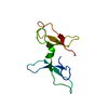 7omkC 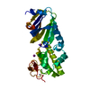 3w0fS S: Starting model for refinement C: citing same article ( |
|---|---|
| Similar structure data |
-Links
 PDBj
PDBj


- Assembly
| Deposited unit | 
| ||||||||
|---|---|---|---|---|---|---|---|---|---|
| 1 |
| ||||||||
| Unit cell |
|
-Components
| #1: Protein | Mass: 33579.699 Da / Num. of mol.: 1 Source method: isolated from a genetically manipulated source Source: (gene. exp.)  References: UniProt: Q8K203, Hydrolases; Glycosylases; Hydrolysing N-glycosyl compounds, DNA-(apurinic or apyrimidinic site) lyase |
|---|---|
| #2: DNA chain | Mass: 5719.699 Da / Num. of mol.: 1 Source method: isolated from a genetically manipulated source Source: (gene. exp.) synthetic construct (others) / Production host: synthetic construct (others) |
| #3: Chemical | ChemComp-ZN /   Mass: 65.409 Da / Num. of mol.: 1 / Source method: obtained synthetically / Formula: Zn Mass: 65.409 Da / Num. of mol.: 1 / Source method: obtained synthetically / Formula: Zn |
| #4: Water | ChemComp-HOH /  Mass: 18.015 Da / Num. of mol.: 123 / Source method: isolated from a natural source / Formula: H2O Mass: 18.015 Da / Num. of mol.: 123 / Source method: isolated from a natural source / Formula: H2O |
| Has ligand of interest | Y |
| Has protein modification | Y |
-Experimental details
-Experiment
| Experiment | Method: X-RAY DIFFRACTION / Number of used crystals: 1 |
|---|
- Sample preparation
| Crystal | Density Matthews: 2.41 Å3/Da / Density % sol: 48.99 % |
|---|---|
| Crystal grow | Temperature: 295 K / Method: vapor diffusion, sitting drop Details: 50 mM PIPES pH = 7.5 4% (w/v) PEG 8000 20 mM MgCl2 1 mM Spermine |
-Data collection
| Diffraction | Mean temperature: 100 K / Serial crystal experiment: N |
|---|---|
| Diffraction source | Source: SYNCHROTRON / Site: BESSY  / Beamline: 14.2 / Wavelength: 0.9184 Å / Beamline: 14.2 / Wavelength: 0.9184 Å |
| Detector | Type: DECTRIS PILATUS 6M / Detector: PIXEL / Date: Jun 23, 2021 |
| Radiation | Protocol: SINGLE WAVELENGTH / Monochromatic (M) / Laue (L): M / Scattering type: x-ray |
| Radiation wavelength | Wavelength: 0.9184 Å / Relative weight: 1 |
| Reflection | Resolution: 2.26→36.13 Å / Num. obs: 179306 / % possible obs: 99.96 % / Redundancy: 5.89 % / Biso Wilson estimate: 46.91 Å2 / CC1/2: 0.992 / CC star: 0.998 / Rmerge(I) obs: 0.1904 / Rrim(I) all: 0.2008 / Net I/σ(I): 9.11 |
| Reflection shell | Resolution: 2.26→2.341 Å / Rmerge(I) obs: 0.32 / Num. unique obs: 1800 / CC1/2: 0.957 |
- Processing
| Software |
| |||||||||||||||||||||||||||||||||||||||||||||||||
|---|---|---|---|---|---|---|---|---|---|---|---|---|---|---|---|---|---|---|---|---|---|---|---|---|---|---|---|---|---|---|---|---|---|---|---|---|---|---|---|---|---|---|---|---|---|---|---|---|---|---|
| Refinement | Method to determine structure: MOLECULAR REPLACEMENT Starting model: 3W0F Resolution: 2.28→36.13 Å / SU ML: 0.31 / Cross valid method: THROUGHOUT / σ(F): 1.34 / Phase error: 27.8 / Stereochemistry target values: ML
| |||||||||||||||||||||||||||||||||||||||||||||||||
| Solvent computation | Shrinkage radii: 0.9 Å / VDW probe radii: 1.11 Å / Solvent model: FLAT BULK SOLVENT MODEL | |||||||||||||||||||||||||||||||||||||||||||||||||
| Displacement parameters | Biso max: 118.98 Å2 / Biso mean: 41.8284 Å2 / Biso min: 18.21 Å2 | |||||||||||||||||||||||||||||||||||||||||||||||||
| Refinement step | Cycle: final / Resolution: 2.28→36.13 Å
| |||||||||||||||||||||||||||||||||||||||||||||||||
| LS refinement shell | Refine-ID: X-RAY DIFFRACTION / Rfactor Rfree error: 0 / Total num. of bins used: 6
|
