 Movie
Movie Controller


+ Open data

- Basic information
| Entry | Database: PDB / ID: 7z2x | ||||||
|---|---|---|---|---|---|---|---|
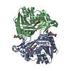
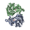
| Title | Wild-type ferulic acid esterase from Lactobacillus buchneri | ||||||
 Components Components | Ferulic acid esterase | ||||||
 Keywords Keywords | HYDROLASE / Ferulic acid esterase / Lactobacillus buchneri / wild type | ||||||
| Function / homology | : / feruloyl esterase / feruloyl esterase activity / Serine aminopeptidase, S33 / Serine aminopeptidase, S33 / Alpha/Beta hydrolase fold / metal ion binding / Ferulic acid esterase Function and homology information Function and homology information | ||||||
| Biological species |  Lentilactobacillus buchneri (bacteria) Lentilactobacillus buchneri (bacteria) | ||||||
| Method |  X-RAY DIFFRACTION / SYNCHROTRON / FOURIER SYNTHESIS / Resolution: 1.5 Å X-RAY DIFFRACTION / SYNCHROTRON / FOURIER SYNTHESIS / Resolution: 1.5 Å | ||||||
 Authors Authors | Mogodiniyai, K.K. / Reichenbach, T. / Kalyani, D.C. / Keskitalo, M.M. / Divne, C. | ||||||
| Funding support |  Sweden, 1items Sweden, 1items
| ||||||
 Citation Citation | Journal: Front Microbiol / Year: 2022 Title: Crystal structure of the feruloyl esterase from Lentilactobacillus buchneri reveals a novel homodimeric state. Authors: Kasmaei, K.M. / Kalyani, D.C. / Reichenbach, T. / Jimenez-Quero, A. / Vilaplana, F. / Divne, C. | ||||||
| History |
|
- Structure visualization
| Structure viewer | Molecule: MolmilJmol/JSmol |
|---|
- Downloads & links
-Download
| PDBx/mmCIF format | 7z2x.cif.gz | 233.3 KB | Display | PDBx/mmCIF format |
|---|---|---|---|---|
| PDB format | pdb7z2x.ent.gz | 185.7 KB | Display | PDB format |
| PDBx/mmJSON format | 7z2x.json.gz | Tree view | PDBx/mmJSON format | |
| Others |  Other downloads Other downloads |
-Validation report
| Arichive directory | https://data.pdbj.org/pub/pdb/validation_reports/z2/7z2xftp://data.pdbj.org/pub/pdb/validation_reports/z2/7z2x | HTTPS FTP |
|---|
-Related structure data
-Links
 PDBj
PDBj- Assembly
| Deposited unit | 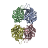
| ||||||||
|---|---|---|---|---|---|---|---|---|---|
| 1 | 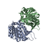
| ||||||||
| 2 | 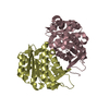
| ||||||||
| Unit cell |
|
-Components
| #1: Protein | Mass: 31690.588 Da / Num. of mol.: 4 Source method: isolated from a genetically manipulated source Source: (gene. exp.) Lentilactobacillus buchneri (bacteria) / Gene: faeA / Plasmid: pNIC28-Bsa4 / Production host: #2: Water | ChemComp-HOH / |  Mass: 18.015 Da / Num. of mol.: 879 / Source method: isolated from a natural source / Formula: H2O Mass: 18.015 Da / Num. of mol.: 879 / Source method: isolated from a natural source / Formula: H2O |
|---|
-Experimental details
-Experiment
| Experiment | Method: X-RAY DIFFRACTION / Number of used crystals: 1 |
|---|
- Sample preparation
| Crystal | Density Matthews: 2.28 Å3/Da / Density % sol: 46.02 % |
|---|---|
| Crystal grow | Temperature: 295 K / Method: vapor diffusion, sitting drop / Details: 0.1 M Bis-Tris pH 5.5, 0.2 M NaCl, 25% P3350 |
-Data collection
| Diffraction | Mean temperature: 100 K / Serial crystal experiment: N |
|---|---|
| Diffraction source | Source: SYNCHROTRON / Site: Diamond  / Beamline: I24 / Wavelength: 0.96852 Å / Beamline: I24 / Wavelength: 0.96852 Å |
| Detector | Type: DECTRIS PILATUS3 6M / Detector: PIXEL / Date: Feb 15, 2020 |
| Radiation | Protocol: SINGLE WAVELENGTH / Monochromatic (M) / Laue (L): M / Scattering type: x-ray |
| Radiation wavelength | Wavelength: 0.96852 Å / Relative weight: 1 |
| Reflection | Resolution: 1.5→30.934 Å / Num. obs: 155659 / % possible obs: 93.5 % / Redundancy: 2.8 % / Biso Wilson estimate: 14.4 Å2 / CC1/2: 0.988 / Net I/σ(I): 6.7 |
| Reflection shell | Resolution: 1.5→1.6 Å / Mean I/σ(I) obs: 2.3 / Num. unique obs: 26396 / CC1/2: 0.528 / % possible all: 90 |
- Processing
| Software |
| |||||||||||||||||||||||||||||||||||||||||||||||||||||||||||||||||||||||||||||||||||||||||||||||||||||||||
|---|---|---|---|---|---|---|---|---|---|---|---|---|---|---|---|---|---|---|---|---|---|---|---|---|---|---|---|---|---|---|---|---|---|---|---|---|---|---|---|---|---|---|---|---|---|---|---|---|---|---|---|---|---|---|---|---|---|---|---|---|---|---|---|---|---|---|---|---|---|---|---|---|---|---|---|---|---|---|---|---|---|---|---|---|---|---|---|---|---|---|---|---|---|---|---|---|---|---|---|---|---|---|---|---|---|---|
| Refinement | Method to determine structure: FOURIER SYNTHESIS Starting model: L. buchneri FAE Resolution: 1.5→30.934 Å / SU ML: 0.19 / Cross valid method: FREE R-VALUE / σ(F): 1.96 / Phase error: 22 / Stereochemistry target values: ML
| |||||||||||||||||||||||||||||||||||||||||||||||||||||||||||||||||||||||||||||||||||||||||||||||||||||||||
| Solvent computation | Shrinkage radii: 0.9 Å / VDW probe radii: 1.11 Å / Solvent model: FLAT BULK SOLVENT MODEL | |||||||||||||||||||||||||||||||||||||||||||||||||||||||||||||||||||||||||||||||||||||||||||||||||||||||||
| Refinement step | Cycle: LAST / Resolution: 1.5→30.934 Å
| |||||||||||||||||||||||||||||||||||||||||||||||||||||||||||||||||||||||||||||||||||||||||||||||||||||||||
| Refine LS restraints |
| |||||||||||||||||||||||||||||||||||||||||||||||||||||||||||||||||||||||||||||||||||||||||||||||||||||||||
| LS refinement shell |
|
