 Movie
Movie Controller

[English] 日本語
 Yorodumi
- PDB-7z2r: Differences between the GluD1 and GluD2 receptors revealed by Glu... -
+ Open data

- Basic information
| Entry | Database: PDB / ID: 7z2r | ||||||
|---|---|---|---|---|---|---|---|
| Title | Differences between the GluD1 and GluD2 receptors revealed by GluD1 X-ray crystallography, binding studies and molecular dynamics | ||||||
 Components Components | Glutamate receptor ionotropic, delta-1 | ||||||
 Keywords Keywords | MEMBRANE PROTEIN / GLUD1 / IONOTROPIC GLUTAMATE RECEPTOR / LIGAND-BINDING DOMAIN / SIGNALLING PROTEIN / DELTA-1 / APO / STRUCTURAL PROTEIN | ||||||
| Function / homology |  Function and homology information Function and homology informationGABA receptor activity / trans-synaptic protein complex / negative regulation of synaptic plasticity / G protein-coupled receptor activity involved in regulation of postsynaptic membrane potential / synaptic signaling via neuropeptide / regulation of postsynapse organization / AMPA glutamate receptor activity / AMPA glutamate receptor complex / social behavior / regulation of postsynaptic membrane neurotransmitter receptor levels ...GABA receptor activity / trans-synaptic protein complex / negative regulation of synaptic plasticity / G protein-coupled receptor activity involved in regulation of postsynaptic membrane potential / synaptic signaling via neuropeptide / regulation of postsynapse organization / AMPA glutamate receptor activity / AMPA glutamate receptor complex / social behavior / regulation of postsynaptic membrane neurotransmitter receptor levels / synaptic transmission, glutamatergic / transmitter-gated monoatomic ion channel activity involved in regulation of postsynaptic membrane potential / postsynaptic density membrane / modulation of chemical synaptic transmission / GABA-ergic synapse / phospholipase C-activating G protein-coupled receptor signaling pathway / dendritic spine / postsynaptic membrane / glutamatergic synapse / metal ion binding / identical protein binding / plasma membrane Similarity search - Function | ||||||
| Biological species |  | ||||||
| Method |  X-RAY DIFFRACTION / SYNCHROTRON / MOLECULAR REPLACEMENT / Resolution: 2.574 Å X-RAY DIFFRACTION / SYNCHROTRON / MOLECULAR REPLACEMENT / Resolution: 2.574 Å | ||||||
 Authors Authors | Masternak, M. / Laulumaa, S. / Kastrup, J.S. | ||||||
| Funding support |  Denmark, 1items Denmark, 1items
| ||||||
 Citation Citation | Journal: Febs J. / Year: 2023 Title: Differences between the GluD1 and GluD2 receptors revealed by GluD1 X-ray crystallography, binding studies and molecular dynamics. Authors: Masternak, M. / Koch, A. / Laulumaa, S. / Tapken, D. / Hollmann, M. / Jorgensen, F.S. / Kastrup, J.S. | ||||||
| History |
|
- Structure visualization
| Structure viewer | Molecule: MolmilJmol/JSmol |
|---|
- Downloads & links
-Download
| PDBx/mmCIF format | 7z2r.cif.gz | 301.8 KB | Display | PDBx/mmCIF format |
|---|---|---|---|---|
| PDB format | pdb7z2r.ent.gz | 251 KB | Display | PDB format |
| PDBx/mmJSON format | 7z2r.json.gz | Tree view | PDBx/mmJSON format | |
| Others |  Other downloads Other downloads |
-Validation report
| Arichive directory | https://data.pdbj.org/pub/pdb/validation_reports/z2/7z2rftp://data.pdbj.org/pub/pdb/validation_reports/z2/7z2r | HTTPS FTP |
|---|
-Related structure data
| Related structure data |  2v3tS S: Starting model for refinement |
|---|---|
| Similar structure data |
-Links
 PDBj
PDBj

- Assembly
| Deposited unit | 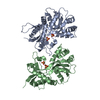
| ||||||||||||||||||||||||||||||||||||||||||||||||||||||||||||||||||||||||||||||||||||||
|---|---|---|---|---|---|---|---|---|---|---|---|---|---|---|---|---|---|---|---|---|---|---|---|---|---|---|---|---|---|---|---|---|---|---|---|---|---|---|---|---|---|---|---|---|---|---|---|---|---|---|---|---|---|---|---|---|---|---|---|---|---|---|---|---|---|---|---|---|---|---|---|---|---|---|---|---|---|---|---|---|---|---|---|---|---|---|---|
| 1 | 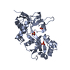
| ||||||||||||||||||||||||||||||||||||||||||||||||||||||||||||||||||||||||||||||||||||||
| 2 | 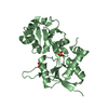
| ||||||||||||||||||||||||||||||||||||||||||||||||||||||||||||||||||||||||||||||||||||||
| Unit cell |
| ||||||||||||||||||||||||||||||||||||||||||||||||||||||||||||||||||||||||||||||||||||||
| Noncrystallographic symmetry (NCS) | NCS domain:
NCS domain segments: Ens-ID: 1 / Beg auth comp-ID: GLY / Beg label comp-ID: GLY
|