Movie
Movie Controller


+ Open data

- Basic information
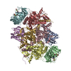
| Entry | Database: PDB / ID: 7z19 | ||||||
|---|---|---|---|---|---|---|---|
| Title | E. coli C-P lyase bound to a single PhnK ABC domain | ||||||
 Components Components |
| ||||||
 Keywords Keywords | TRANSFERASE / protein complex / ABC / hydrolase / lyase / carbon phosphorus | ||||||
| Function / homology |  Function and homology information Function and homology informationalpha-D-ribose 1-methylphosphonate 5-phosphate C-P-lyase / alpha-D-ribose 1-methylphosphonate 5-phosphate C-P-lyase activity / alpha-D-ribose 1-methylphosphonate 5-triphosphate synthase / alpha-D-ribose 1-methylphosphonate 5-triphosphate synthase activity / alpha-D-ribose 1-methylphosphonate 5-triphosphate synthase complex / carbon phosphorus lyase complex / organic phosphonate metabolic process / organic phosphonate transport / organic phosphonate catabolic process / peptide transport ...alpha-D-ribose 1-methylphosphonate 5-phosphate C-P-lyase / alpha-D-ribose 1-methylphosphonate 5-phosphate C-P-lyase activity / alpha-D-ribose 1-methylphosphonate 5-triphosphate synthase / alpha-D-ribose 1-methylphosphonate 5-triphosphate synthase activity / alpha-D-ribose 1-methylphosphonate 5-triphosphate synthase complex / carbon phosphorus lyase complex / organic phosphonate metabolic process / organic phosphonate transport / organic phosphonate catabolic process / peptide transport / 4 iron, 4 sulfur cluster binding / lyase activity / protein homodimerization activity / ATP hydrolysis activity / ATP binding / metal ion binding / identical protein binding Similarity search - Function | ||||||
| Biological species |  | ||||||
| Method | ELECTRON MICROSCOPY / single particle reconstruction / cryo EM / Resolution: 2.57 Å | ||||||
 Authors Authors | Amstrup, S.K. / Sofos, N. / Karlsen, J.L. / Skjerning, R.B. / Boesen, T. / Enghild, J.J. / Hove-Jensen, B. / Brodersen, D.E. | ||||||
| Funding support |  Denmark, 1items Denmark, 1items
| ||||||
 Citation Citation | Journal: Nat Commun / Year: 2023 Title: Structural remodelling of the carbon-phosphorus lyase machinery by a dual ABC ATPase. Authors: Søren K Amstrup / Sui Ching Ong / Nicholas Sofos / Jesper L Karlsen / Ragnhild B Skjerning / Thomas Boesen / Jan J Enghild / Bjarne Hove-Jensen / Ditlev E Brodersen / Abstract: In Escherichia coli, the 14-cistron phn operon encoding carbon-phosphorus lyase allows for utilisation of phosphorus from a wide range of stable phosphonate compounds containing a C-P bond. As part ...In Escherichia coli, the 14-cistron phn operon encoding carbon-phosphorus lyase allows for utilisation of phosphorus from a wide range of stable phosphonate compounds containing a C-P bond. As part of a complex, multi-step pathway, the PhnJ subunit was shown to cleave the C-P bond via a radical mechanism, however, the details of the reaction could not immediately be reconciled with the crystal structure of a 220 kDa PhnGHIJ C-P lyase core complex, leaving a significant gap in our understanding of phosphonate breakdown in bacteria. Here, we show using single-particle cryogenic electron microscopy that PhnJ mediates binding of a double dimer of the ATP-binding cassette proteins, PhnK and PhnL, to the core complex. ATP hydrolysis induces drastic structural remodelling leading to opening of the core complex and reconfiguration of a metal-binding and putative active site located at the interface between the PhnI and PhnJ subunits. #1: Journal: Biorxiv / Year: 2022Title: Structural remodelling of the carbon-phosphorus lyase machinery by a dual ABC ATPase Authors: Amstrup, S.K. / Sofos, N. / Karlsen, J.L. / Skjerning, R.B. / Boesen, T. / Enghild, J.J. / Hove-Jensen, B. / Brodersen, D.E. | ||||||
| History |
|
- Structure visualization
| Structure viewer | Molecule: MolmilJmol/JSmol |
|---|
- Downloads & links
-Download
| PDBx/mmCIF format | 7z19.cif.gz | 705.3 KB | Display | PDBx/mmCIF format |
|---|---|---|---|---|
| PDB format | pdb7z19.ent.gz | 586.4 KB | Display | PDB format |
| PDBx/mmJSON format | 7z19.json.gz | Tree view | PDBx/mmJSON format | |
| Others |  Other downloads Other downloads |
-Validation report
| Arichive directory | https://data.pdbj.org/pub/pdb/validation_reports/z1/7z19ftp://data.pdbj.org/pub/pdb/validation_reports/z1/7z19 | HTTPS FTP |
|---|
-Related structure data
| Related structure data |  14445MC 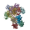 7z15C 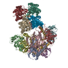 7z16C 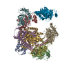 7z17C 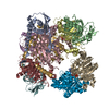 7z18C M: map data used to model this data C: citing same article ( |
|---|---|
| Similar structure data |
-Links
 PDBj
PDBj

- Assembly
| Deposited unit | 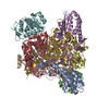
|
|---|---|
| 1 |
|
-Components
-Alpha-D-ribose 1-methylphosphonate 5-triphosphate synthase subunit ... , 3 types, 6 molecules AEBFCG
| #1: Protein | Mass: 16560.637 Da / Num. of mol.: 2 Source method: isolated from a genetically manipulated source Details: pBW120 / Source: (gene. exp.) References: UniProt: P16685, alpha-D-ribose 1-methylphosphonate 5-triphosphate synthase #2: Protein | Mass: 21074.271 Da / Num. of mol.: 2 Source method: isolated from a genetically manipulated source Details: pBW120 / Source: (gene. exp.) References: UniProt: P16686, alpha-D-ribose 1-methylphosphonate 5-triphosphate synthase #3: Protein | Mass: 38922.707 Da / Num. of mol.: 2 Source method: isolated from a genetically manipulated source Source: (gene. exp.) References: UniProt: P16687, alpha-D-ribose 1-methylphosphonate 5-triphosphate synthase |
|---|
-Protein , 2 types, 3 molecules DHI
| #4: Protein | Mass: 31879.088 Da / Num. of mol.: 2 Source method: isolated from a genetically manipulated source Source: (gene. exp.) References: UniProt: P16688, alpha-D-ribose 1-methylphosphonate 5-phosphate C-P-lyase #5: Protein | | Mass: 28665.652 Da / Num. of mol.: 1 Source method: isolated from a genetically manipulated source Source: (gene. exp.) |
|---|
-Non-polymers , 3 types, 483 molecules 


| #6: Chemical | ChemComp-ZN /  Mass: 65.409 Da / Num. of mol.: 4 / Source method: obtained synthetically / Formula: Zn Mass: 65.409 Da / Num. of mol.: 4 / Source method: obtained synthetically / Formula: Zn#7: Chemical |  Mass: 94.971 Da / Num. of mol.: 2 / Source method: obtained synthetically / Formula: PO4 Mass: 94.971 Da / Num. of mol.: 2 / Source method: obtained synthetically / Formula: PO4#8: Water | ChemComp-HOH / | Mass: 18.015 Da / Num. of mol.: 477 / Source method: isolated from a natural source / Formula: H2O |
|---|
-Details
| Has ligand of interest | N |
|---|
-Experimental details
-Experiment
| Experiment | Method: ELECTRON MICROSCOPY |
|---|---|
| EM experiment | Aggregation state: PARTICLE / 3D reconstruction method: single particle reconstruction |
- Sample preparation
| Component | Name: E. coli C-P lyase bound to a single PhnK ABC domain / Type: COMPLEX Details: Complex of Phn(GHIJ)2K with a single PhnK subunit bound to the PhnGHIJ core complex Entity ID: #1-#5 / Source: RECOMBINANT | ||||||||||||||||||||
|---|---|---|---|---|---|---|---|---|---|---|---|---|---|---|---|---|---|---|---|---|---|
| Molecular weight | Value: 0.252 MDa / Experimental value: YES | ||||||||||||||||||||
| Source (natural) | Organism: | ||||||||||||||||||||
| Source (recombinant) | Organism: | ||||||||||||||||||||
| Buffer solution | pH: 7.5 | ||||||||||||||||||||
| Buffer component |
| ||||||||||||||||||||
| Specimen | Conc.: 1.7 mg/ml / Embedding applied: NO / Shadowing applied: NO / Staining applied: NO / Vitrification applied: YES / Details: Sample was monodisperse. | ||||||||||||||||||||
| Specimen support | Details: 15 mA / Grid material: COPPER / Grid mesh size: 400 divisions/in. / Grid type: C-flat-1.2/1.3 | ||||||||||||||||||||
| Vitrification | Instrument: LEICA EM GP / Cryogen name: ETHANE / Humidity: 100 % / Chamber temperature: 278 K / Details: Blotting for 6-9 seconds before plunging |
- Electron microscopy imaging
| Experimental equipment |  Model: Titan Krios / Image courtesy: FEI Company |
|---|---|
| Microscopy | Model: FEI TITAN KRIOS |
| Electron gun | Electron source:  FIELD EMISSION GUN / Accelerating voltage: 300 kV / Illumination mode: FLOOD BEAM FIELD EMISSION GUN / Accelerating voltage: 300 kV / Illumination mode: FLOOD BEAM |
| Electron lens | Mode: BRIGHT FIELD / Nominal magnification: 135000 X / Nominal defocus max: 2000 nm / Nominal defocus min: 700 nm / Cs: 2.7 mm / Alignment procedure: COMA FREE |
| Image recording | Electron dose: 52 e/Å2 / Film or detector model: GATAN K3 (6k x 4k) / Num. of grids imaged: 1 / Num. of real images: 17088 Details: Images were collected in movie-mode with 0.2 seconds per frame with a total of 40 frames |
| EM imaging optics | Energyfilter slit width: 20 eV |
- Processing
| EM software |
| |||||||||||||||||||||||||||||||||||||||||||||
|---|---|---|---|---|---|---|---|---|---|---|---|---|---|---|---|---|---|---|---|---|---|---|---|---|---|---|---|---|---|---|---|---|---|---|---|---|---|---|---|---|---|---|---|---|---|---|
| CTF correction | Type: PHASE FLIPPING AND AMPLITUDE CORRECTION | |||||||||||||||||||||||||||||||||||||||||||||
| Particle selection | Num. of particles selected: 20000000 / Details: LoG picker from relion.py | |||||||||||||||||||||||||||||||||||||||||||||
| Symmetry | Point symmetry: C1 (asymmetric) | |||||||||||||||||||||||||||||||||||||||||||||
| 3D reconstruction | Resolution: 2.57 Å / Resolution method: FSC 0.143 CUT-OFF / Num. of particles: 50323 / Algorithm: BACK PROJECTION / Num. of class averages: 1 / Symmetry type: POINT | |||||||||||||||||||||||||||||||||||||||||||||
| Atomic model building | B value: 54.2 / Space: RECIPROCAL | |||||||||||||||||||||||||||||||||||||||||||||
| Atomic model building | PDB-ID: 4XB6 Accession code: 4XB6 / Source name: PDB / Type: experimental model |