 ムービー
ムービー コントローラー


+ データを開く

- 基本情報
基本情報
| 登録情報 | データベース: PDB / ID: 7z0t | ||||||
|---|---|---|---|---|---|---|---|
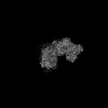
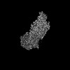
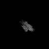
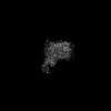
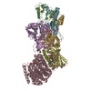
| タイトル | Structure of the Escherichia coli formate hydrogenlyase complex (aerobic preparation, composite structure) | ||||||
 要素 要素 |
| ||||||
 キーワード キーワード | MEMBRANE PROTEIN / FHL / group-4 membrane bound hydrogenase / [NiFe] hydrogenase | ||||||
| 機能・相同性 |  機能・相同性情報 機能・相同性情報formate dehydrogenase (hydrogenase) / formate oxidation / oxidoreductase activity, acting on the aldehyde or oxo group of donors / formate dehydrogenase complex / glucose catabolic process / formate dehydrogenase (NAD+) activity / anaerobic electron transport chain / urate catabolic process / molybdopterin cofactor binding / anaerobic respiration ...formate dehydrogenase (hydrogenase) / formate oxidation / oxidoreductase activity, acting on the aldehyde or oxo group of donors / formate dehydrogenase complex / glucose catabolic process / formate dehydrogenase (NAD+) activity / anaerobic electron transport chain / urate catabolic process / molybdopterin cofactor binding / anaerobic respiration / nickel cation binding / oxidoreductase activity, acting on NAD(P)H / NADH dehydrogenase (ubiquinone) activity / quinone binding / ATP synthesis coupled electron transport / aerobic respiration / respiratory electron transport chain / NAD binding / 4 iron, 4 sulfur cluster binding / oxidoreductase activity / membrane / metal ion binding / plasma membrane / cytosol 類似検索 - 分子機能 | ||||||
| 生物種 |  | ||||||
| 手法 | 電子顕微鏡法 / 単粒子再構成法 / クライオ電子顕微鏡法 / 解像度: 3.4 Å | ||||||
 データ登録者 データ登録者 | Steinhilper, R. / Murphy, B.J. | ||||||
| 資金援助 |  ドイツ, 1件 ドイツ, 1件
| ||||||
 引用 引用 | ジャーナル: Nat Commun / 年: 2022 タイトル: Structure of the membrane-bound formate hydrogenlyase complex from Escherichia coli. 著者: Ralf Steinhilper / Gabriele Höff / Johann Heider / Bonnie J Murphy / 要旨: The prototypical hydrogen-producing enzyme, the membrane-bound formate hydrogenlyase (FHL) complex from Escherichia coli, links formate oxidation at a molybdopterin-containing formate dehydrogenase ...The prototypical hydrogen-producing enzyme, the membrane-bound formate hydrogenlyase (FHL) complex from Escherichia coli, links formate oxidation at a molybdopterin-containing formate dehydrogenase to proton reduction at a [NiFe] hydrogenase. It is of intense interest due to its ability to efficiently produce H during fermentation, its reversibility, allowing H-dependent CO reduction, and its evolutionary link to respiratory complex I. FHL has been studied for over a century, but its atomic structure remains unknown. Here we report cryo-EM structures of FHL in its aerobically and anaerobically isolated forms at resolutions reaching 2.6 Å. This includes well-resolved density for conserved loops linking the soluble and membrane arms believed to be essential in coupling enzymatic turnover to ion translocation across the membrane in the complex I superfamily. We evaluate possible structural determinants of the bias toward hydrogen production over its oxidation and describe an unpredicted metal-binding site near the interface of FdhF and HycF subunits that may play a role in redox-dependent regulation of FdhF interaction with the complex. | ||||||
| 履歴 |
|
- 構造の表示
構造の表示
| 構造ビューア | 分子: MolmilJmol/JSmol |
|---|
- ダウンロードとリンク
-ダウンロード
| PDBx/mmCIF形式 | 7z0t.cif.gz | 471.9 KB | 表示 | PDBx/mmCIF形式 |
|---|---|---|---|---|
| PDB形式 | pdb7z0t.ent.gz | 376.3 KB | 表示 | PDB形式 |
| PDBx/mmJSON形式 | 7z0t.json.gz | ツリー表示 | PDBx/mmJSON形式 | |
| その他 |  その他のダウンロード その他のダウンロード |
-検証レポート
| アーカイブディレクトリ | https://data.pdbj.org/pub/pdb/validation_reports/z0/7z0tftp://data.pdbj.org/pub/pdb/validation_reports/z0/7z0t | HTTPS FTP |
|---|
-関連構造データ


-リンク
 PDBj
PDBj


- 集合体
集合体
| 登録構造単位 | 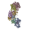
|
|---|---|
| 1 |
|
-要素
-Formate hydrogenlyase subunit ... , 6種, 6分子 CEBGFD
| #1: タンパク質 | 分子量: 64121.742 Da / 分子数: 1 / 由来タイプ: 天然 / 由来: (天然) |
|---|---|
| #2: タンパク質 | 分子量: 66589.859 Da / 分子数: 1 / Mutation: internal deca-His-Gly-Ser sequence after Gly83 / 由来タイプ: 天然 / 由来: (天然) |
| #3: タンパク質 | 分子量: 21899.289 Da / 分子数: 1 / 由来タイプ: 天然 / 由来: (天然) |
| #4: タンパク質 | 分子量: 28033.170 Da / 分子数: 1 / 由来タイプ: 天然 / 由来: (天然) |
| #5: タンパク質 | 分子量: 20336.395 Da / 分子数: 1 / 由来タイプ: 天然 / 由来: (天然) |
| #6: タンパク質 | 分子量: 33049.152 Da / 分子数: 1 / 由来タイプ: 天然 / 由来: (天然) |
-タンパク質 , 1種, 1分子 A
| #7: タンパク質 | 分子量: 79465.703 Da / 分子数: 1 / 由来タイプ: 天然 / 由来: (天然) 参照: UniProt: P07658, formate dehydrogenase (hydrogenase) |
|---|
-非ポリマー , 6種, 14分子 


| #8: 化合物 | ChemComp-NI /  分子量: 58.693 Da / 分子数: 1 / 由来タイプ: 合成 / 式: Ni / タイプ: SUBJECT OF INVESTIGATION 分子量: 58.693 Da / 分子数: 1 / 由来タイプ: 合成 / 式: Ni / タイプ: SUBJECT OF INVESTIGATION | ||||||
|---|---|---|---|---|---|---|---|
| #9: 化合物 | ChemComp-FCO /  分子量: 135.890 Da / 分子数: 1 / 由来タイプ: 合成 / 式: C3FeN2O / タイプ: SUBJECT OF INVESTIGATION 分子量: 135.890 Da / 分子数: 1 / 由来タイプ: 合成 / 式: C3FeN2O / タイプ: SUBJECT OF INVESTIGATION | ||||||
| #10: 化合物 | ChemComp-SF4 /  分子量: 351.640 Da / 分子数: 8 / 由来タイプ: 合成 / 式: Fe4S4 / タイプ: SUBJECT OF INVESTIGATION 分子量: 351.640 Da / 分子数: 8 / 由来タイプ: 合成 / 式: Fe4S4 / タイプ: SUBJECT OF INVESTIGATION#11: 化合物 | ChemComp-FE / |  分子量: 55.845 Da / 分子数: 1 / 由来タイプ: 合成 / 式: Fe / タイプ: SUBJECT OF INVESTIGATION 分子量: 55.845 Da / 分子数: 1 / 由来タイプ: 合成 / 式: Fe / タイプ: SUBJECT OF INVESTIGATION#12: 化合物 |  分子量: 740.557 Da / 分子数: 2 / 由来タイプ: 合成 / 式: C20H26N10O13P2S2 / タイプ: SUBJECT OF INVESTIGATION 分子量: 740.557 Da / 分子数: 2 / 由来タイプ: 合成 / 式: C20H26N10O13P2S2 / タイプ: SUBJECT OF INVESTIGATION#13: 化合物 | ChemComp-6MO / |  分子量: 95.940 Da / 分子数: 1 / 由来タイプ: 合成 / 式: Mo / タイプ: SUBJECT OF INVESTIGATION 分子量: 95.940 Da / 分子数: 1 / 由来タイプ: 合成 / 式: Mo / タイプ: SUBJECT OF INVESTIGATION |
-詳細
| 研究の焦点であるリガンドがあるか | Y |
|---|
-実験情報
-実験
| 実験 | 手法: 電子顕微鏡法 |
|---|---|
| EM実験 | 試料の集合状態: PARTICLE / 3次元再構成法: 単粒子再構成法 |
- 試料調製
試料調製
| 構成要素 | 名称: Escherichia coli formate hydrogenlyase complex / タイプ: COMPLEX / Entity ID: #1-#7 / 由来: NATURAL |
|---|---|
| 由来(天然) | 生物種: |
| 緩衝液 | pH: 7.5 |
| 試料 | 包埋: NO / シャドウイング: NO / 染色: NO / 凍結: YES |
| 試料支持 | グリッドの材料: COPPER / グリッドのタイプ: C-flat-2/1 |
| 急速凍結 | 凍結剤: ETHANE |
- 電子顕微鏡撮影
| 実験機器 |  モデル: Titan Krios / 画像提供: FEI Company |
|---|---|
| 顕微鏡 | モデル: FEI TITAN KRIOS |
| 電子銃 | 電子線源:  FIELD EMISSION GUN / 加速電圧: 300 kV / 照射モード: FLOOD BEAM FIELD EMISSION GUN / 加速電圧: 300 kV / 照射モード: FLOOD BEAM |
| 電子レンズ | モード: BRIGHT FIELD / 最大 デフォーカス(公称値): 2200 nm / 最小 デフォーカス(公称値): 1600 nm / Cs: 2.7 mm |
| 試料ホルダ | 試料ホルダーモデル: FEI TITAN KRIOS AUTOGRID HOLDER |
| 撮影 | 電子線照射量: 72 e/Å2 / 検出モード: COUNTING フィルム・検出器のモデル: GATAN K2 SUMMIT (4k x 4k) |
- 解析
解析
| ソフトウェア | 名称: PHENIX / バージョン: 1.19.2_4158: / 分類: 精密化 | ||||||||||||||||||||||||||||||||
|---|---|---|---|---|---|---|---|---|---|---|---|---|---|---|---|---|---|---|---|---|---|---|---|---|---|---|---|---|---|---|---|---|---|
| EMソフトウェア |
| ||||||||||||||||||||||||||||||||
| CTF補正 | タイプ: PHASE FLIPPING AND AMPLITUDE CORRECTION | ||||||||||||||||||||||||||||||||
| 3次元再構成 | 解像度: 3.4 Å / 解像度の算出法: OTHER / 粒子像の数: 90459 詳細: This is a composite map. The maps that constitute this composite map have resolutions between 3.0 and 3.4 A. These have all been determined by FSC 0.143 cut-off. 対称性のタイプ: POINT | ||||||||||||||||||||||||||||||||
| 拘束条件 |
|
