 Movie
Movie Controller

[English] 日本語
 Yorodumi
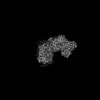
- PDB-7z0s: Structure of the Escherichia coli formate hydrogenlyase complex (... -
+ Open data

- Basic information
| Entry | Database: PDB / ID: 7z0s | ||||||
|---|---|---|---|---|---|---|---|
| Title | Structure of the Escherichia coli formate hydrogenlyase complex (anaerobic preparation, without formate dehydrogenase H) | ||||||
 Components Components | (Formate hydrogenlyase subunit ...) x 6 | ||||||
 Keywords Keywords | MEMBRANE PROTEIN / FHL / group-4 membrane bound hydrogenase / [NiFe] hydrogenase | ||||||
| Function / homology |  Function and homology information Function and homology informationformate oxidation / formate dehydrogenase complex / glucose catabolic process / anaerobic electron transport chain / anaerobic respiration / nickel cation binding / oxidoreductase activity, acting on NAD(P)H / NADH dehydrogenase (ubiquinone) activity / quinone binding / ATP synthesis coupled electron transport ...formate oxidation / formate dehydrogenase complex / glucose catabolic process / anaerobic electron transport chain / anaerobic respiration / nickel cation binding / oxidoreductase activity, acting on NAD(P)H / NADH dehydrogenase (ubiquinone) activity / quinone binding / ATP synthesis coupled electron transport / aerobic respiration / NAD binding / 4 iron, 4 sulfur cluster binding / oxidoreductase activity / membrane / metal ion binding / plasma membrane Similarity search - Function | ||||||
| Biological species |  | ||||||
| Method | ELECTRON MICROSCOPY / single particle reconstruction / cryo EM / Resolution: 2.6 Å | ||||||
 Authors Authors | Steinhilper, R. / Murphy, B.J. | ||||||
| Funding support |  Germany, 1items Germany, 1items
| ||||||
 Citation Citation | Journal: Nat Commun / Year: 2022 Title: Structure of the membrane-bound formate hydrogenlyase complex from Escherichia coli. Authors: Ralf Steinhilper / Gabriele Höff / Johann Heider / Bonnie J Murphy / Abstract: The prototypical hydrogen-producing enzyme, the membrane-bound formate hydrogenlyase (FHL) complex from Escherichia coli, links formate oxidation at a molybdopterin-containing formate dehydrogenase ...The prototypical hydrogen-producing enzyme, the membrane-bound formate hydrogenlyase (FHL) complex from Escherichia coli, links formate oxidation at a molybdopterin-containing formate dehydrogenase to proton reduction at a [NiFe] hydrogenase. It is of intense interest due to its ability to efficiently produce H during fermentation, its reversibility, allowing H-dependent CO reduction, and its evolutionary link to respiratory complex I. FHL has been studied for over a century, but its atomic structure remains unknown. Here we report cryo-EM structures of FHL in its aerobically and anaerobically isolated forms at resolutions reaching 2.6 Å. This includes well-resolved density for conserved loops linking the soluble and membrane arms believed to be essential in coupling enzymatic turnover to ion translocation across the membrane in the complex I superfamily. We evaluate possible structural determinants of the bias toward hydrogen production over its oxidation and describe an unpredicted metal-binding site near the interface of FdhF and HycF subunits that may play a role in redox-dependent regulation of FdhF interaction with the complex. | ||||||
| History |
|
- Structure visualization
| Structure viewer | Molecule: MolmilJmol/JSmol |
|---|
- Downloads & links
-Download
| PDBx/mmCIF format | 7z0s.cif.gz | 376.1 KB | Display | PDBx/mmCIF format |
|---|---|---|---|---|
| PDB format | pdb7z0s.ent.gz | 293.9 KB | Display | PDB format |
| PDBx/mmJSON format | 7z0s.json.gz | Tree view | PDBx/mmJSON format | |
| Others |  Other downloads Other downloads |
-Validation report
| Arichive directory | https://data.pdbj.org/pub/pdb/validation_reports/z0/7z0sftp://data.pdbj.org/pub/pdb/validation_reports/z0/7z0s | HTTPS FTP |
|---|
-Related structure data
| Related structure data | 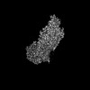 14429MC 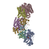 7z0tC M: map data used to model this data C: citing same article ( |
|---|---|
| Similar structure data |


-Links
 PDBj
PDBj


- Assembly
| Deposited unit | 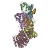
|
|---|---|
| 1 |
|
-Components
-Formate hydrogenlyase subunit ... , 6 types, 6 molecules CEBGFD
| #1: Protein | Mass: 64121.742 Da / Num. of mol.: 1 / Source method: isolated from a natural source / Source: (natural) |
|---|---|
| #2: Protein | Mass: 66589.859 Da / Num. of mol.: 1 / Mutation: internal deca-His-Gly-Ser sequence after Gly83 Source method: isolated from a genetically manipulated source Source: (gene. exp.) |
| #3: Protein | Mass: 21899.289 Da / Num. of mol.: 1 / Source method: isolated from a natural source / Source: (natural) |
| #4: Protein | Mass: 28033.170 Da / Num. of mol.: 1 / Source method: isolated from a natural source / Source: (natural) |
| #5: Protein | Mass: 20336.395 Da / Num. of mol.: 1 / Source method: isolated from a natural source / Source: (natural) |
| #6: Protein | Mass: 33049.152 Da / Num. of mol.: 1 / Source method: isolated from a natural source / Source: (natural) |
-Non-polymers , 9 types, 124 molecules 


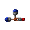
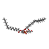


| #7: Chemical | ChemComp-CDL /  Mass: 1464.043 Da / Num. of mol.: 1 / Source method: obtained synthetically / Formula: C81H156O17P2 / Comment: phospholipid*YM Mass: 1464.043 Da / Num. of mol.: 1 / Source method: obtained synthetically / Formula: C81H156O17P2 / Comment: phospholipid*YM | ||||||||||||||
|---|---|---|---|---|---|---|---|---|---|---|---|---|---|---|---|
| #8: Chemical |  Mass: 734.039 Da / Num. of mol.: 2 / Source method: obtained synthetically / Formula: C40H80NO8P / Comment: phospholipid*YM Mass: 734.039 Da / Num. of mol.: 2 / Source method: obtained synthetically / Formula: C40H80NO8P / Comment: phospholipid*YM#9: Chemical | ChemComp-NI / |  Mass: 58.693 Da / Num. of mol.: 1 / Source method: obtained synthetically / Formula: Ni / Feature type: SUBJECT OF INVESTIGATION Mass: 58.693 Da / Num. of mol.: 1 / Source method: obtained synthetically / Formula: Ni / Feature type: SUBJECT OF INVESTIGATION#10: Chemical | ChemComp-FCO / |  Mass: 135.890 Da / Num. of mol.: 1 / Source method: obtained synthetically / Formula: C3FeN2O / Feature type: SUBJECT OF INVESTIGATION Mass: 135.890 Da / Num. of mol.: 1 / Source method: obtained synthetically / Formula: C3FeN2O / Feature type: SUBJECT OF INVESTIGATION#11: Chemical | ChemComp-DR9 / |  Mass: 746.991 Da / Num. of mol.: 1 / Source method: obtained synthetically / Formula: C40H75O10P Mass: 746.991 Da / Num. of mol.: 1 / Source method: obtained synthetically / Formula: C40H75O10P#12: Chemical | ChemComp-SF4 /  Mass: 351.640 Da / Num. of mol.: 7 / Source method: obtained synthetically / Formula: Fe4S4 / Feature type: SUBJECT OF INVESTIGATION Mass: 351.640 Da / Num. of mol.: 7 / Source method: obtained synthetically / Formula: Fe4S4 / Feature type: SUBJECT OF INVESTIGATION#13: Chemical | ChemComp-FE / |  Mass: 55.845 Da / Num. of mol.: 1 / Source method: obtained synthetically / Formula: Fe / Feature type: SUBJECT OF INVESTIGATION Mass: 55.845 Da / Num. of mol.: 1 / Source method: obtained synthetically / Formula: Fe / Feature type: SUBJECT OF INVESTIGATION#14: Chemical | ChemComp-LMN / |  Mass: 1005.188 Da / Num. of mol.: 1 / Source method: obtained synthetically / Formula: C47H88O22 / Comment: detergent*YM Mass: 1005.188 Da / Num. of mol.: 1 / Source method: obtained synthetically / Formula: C47H88O22 / Comment: detergent*YM#15: Water | ChemComp-HOH / | Mass: 18.015 Da / Num. of mol.: 109 / Source method: isolated from a natural source / Formula: H2O |
-Details
| Has ligand of interest | Y |
|---|
-Experimental details
-Experiment
| Experiment | Method: ELECTRON MICROSCOPY |
|---|---|
| EM experiment | Aggregation state: PARTICLE / 3D reconstruction method: single particle reconstruction |
- Sample preparation
| Component | Name: Escherichia coli formate hydrogenlyase complex without formate dehydrogenase H Type: COMPLEX / Details: Sample was prepared anaerobically / Entity ID: #1-#6 / Source: MULTIPLE SOURCES |
|---|---|
| Source (natural) | Organism: |
| Buffer solution | pH: 7.5 |
| Specimen | Embedding applied: NO / Shadowing applied: NO / Staining applied: NO / Vitrification applied: YES |
| Specimen support | Grid material: COPPER / Grid type: C-flat-1.2/1.3 |
| Vitrification | Cryogen name: ETHANE |
- Electron microscopy imaging
| Experimental equipment |  Model: Titan Krios / Image courtesy: FEI Company |
|---|---|
| Microscopy | Model: FEI TITAN KRIOS |
| Electron gun | Electron source:  FIELD EMISSION GUN / Accelerating voltage: 300 kV / Illumination mode: FLOOD BEAM FIELD EMISSION GUN / Accelerating voltage: 300 kV / Illumination mode: FLOOD BEAM |
| Electron lens | Mode: BRIGHT FIELD / Nominal defocus max: 2500 nm / Nominal defocus min: 1000 nm / Cs: 2.7 mm |
| Specimen holder | Specimen holder model: FEI TITAN KRIOS AUTOGRID HOLDER |
| Image recording | Electron dose: 61 e/Å2 / Film or detector model: GATAN K3 BIOQUANTUM (6k x 4k) |
- Processing
| Software | Name: PHENIX / Version: 1.19.2_4158: / Classification: refinement | ||||||||||||||||||||||||||||||||
|---|---|---|---|---|---|---|---|---|---|---|---|---|---|---|---|---|---|---|---|---|---|---|---|---|---|---|---|---|---|---|---|---|---|
| EM software |
| ||||||||||||||||||||||||||||||||
| CTF correction | Type: PHASE FLIPPING AND AMPLITUDE CORRECTION | ||||||||||||||||||||||||||||||||
| 3D reconstruction | Resolution: 2.6 Å / Resolution method: FSC 0.143 CUT-OFF / Num. of particles: 300386 / Symmetry type: POINT | ||||||||||||||||||||||||||||||||
| Refine LS restraints |
|
