 Movie
Movie Controller


+ Open data

- Basic information
| Entry | Database: PDB / ID: 7z00 | ||||||
|---|---|---|---|---|---|---|---|
| Title | Crystal structure of Vibrio alkaline phosphatase in 1.0 M KBr | ||||||
 Components Components | Alkaline phosphatase | ||||||
 Keywords Keywords | HYDROLASE / Alkaline phosphatase / AP | ||||||
| Function / homology |  Function and homology information Function and homology information | ||||||
| Biological species |  Vibrio sp. G15-21 (bacteria) Vibrio sp. G15-21 (bacteria) | ||||||
| Method |  X-RAY DIFFRACTION / SYNCHROTRON / MOLECULAR REPLACEMENT / Resolution: 2.6 Å X-RAY DIFFRACTION / SYNCHROTRON / MOLECULAR REPLACEMENT / Resolution: 2.6 Å | ||||||
 Authors Authors | Markusson, S. / Hjorleifsson, J.G. / Kursula, P. / Asgeirsson, B. | ||||||
| Funding support |  Iceland, 1items Iceland, 1items
| ||||||
 Citation Citation | Journal: Biochemistry / Year: 2022 Title: Structural Characterization of Functionally Important Chloride Binding Sites in the Marine Vibrio Alkaline Phosphatase. Authors: Markusson, S. / Hjorleifsson, J.G. / Kursula, P. / Asgeirsson, B. | ||||||
| History |
|
- Structure visualization
| Structure viewer | Molecule: MolmilJmol/JSmol |
|---|
- Downloads & links
-Download
| PDBx/mmCIF format | 7z00.cif.gz | 227.2 KB | Display | PDBx/mmCIF format |
|---|---|---|---|---|
| PDB format | pdb7z00.ent.gz | 154.5 KB | Display | PDB format |
| PDBx/mmJSON format | 7z00.json.gz | Tree view | PDBx/mmJSON format | |
| Others |  Other downloads Other downloads |
-Validation report
| Arichive directory | https://data.pdbj.org/pub/pdb/validation_reports/z0/7z00ftp://data.pdbj.org/pub/pdb/validation_reports/z0/7z00 | HTTPS FTP |
|---|
-Related structure data
| Related structure data |  7qowC  7qp8C  7yzzC  3e2dS S: Starting model for refinement C: citing same article ( |
|---|---|
| Similar structure data |
-Links
 PDBj
PDBj


- Assembly
| Deposited unit | 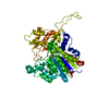
| ||||||||||||
|---|---|---|---|---|---|---|---|---|---|---|---|---|---|
| 1 | 
| ||||||||||||
| Unit cell |
|
-Components
-Protein , 1 types, 1 molecules B
| #1: Protein | Mass: 58616.484 Da / Num. of mol.: 1 Source method: isolated from a genetically manipulated source Source: (gene. exp.) Vibrio sp. G15-21 (bacteria) / Production host: |
|---|
-Non-polymers , 5 types, 9 molecules 


| #2: Chemical |  Mass: 65.409 Da / Num. of mol.: 2 / Source method: obtained synthetically / Formula: Zn Mass: 65.409 Da / Num. of mol.: 2 / Source method: obtained synthetically / Formula: Zn#3: Chemical | ChemComp-BR / |  Mass: 79.904 Da / Num. of mol.: 1 / Source method: obtained synthetically / Formula: Br / Feature type: SUBJECT OF INVESTIGATION Mass: 79.904 Da / Num. of mol.: 1 / Source method: obtained synthetically / Formula: Br / Feature type: SUBJECT OF INVESTIGATION#4: Chemical |  Mass: 24.305 Da / Num. of mol.: 2 / Source method: obtained synthetically / Formula: Mg Mass: 24.305 Da / Num. of mol.: 2 / Source method: obtained synthetically / Formula: Mg#5: Chemical | ChemComp-PO4 / |  Mass: 94.971 Da / Num. of mol.: 1 / Source method: obtained synthetically / Formula: PO4 Mass: 94.971 Da / Num. of mol.: 1 / Source method: obtained synthetically / Formula: PO4#6: Water | ChemComp-HOH / | Mass: 18.015 Da / Num. of mol.: 3 / Source method: isolated from a natural source / Formula: H2O |
|---|
-Details
| Has ligand of interest | Y |
|---|
-Experimental details
-Experiment
| Experiment | Method: X-RAY DIFFRACTION / Number of used crystals: 1 |
|---|
- Sample preparation
| Crystal | Density Matthews: 2.19 Å3/Da / Density % sol: 43.9 % |
|---|---|
| Crystal grow | Temperature: 293 K / Method: vapor diffusion, hanging drop / pH: 7 Details: 13.4 mg/mL protein in 24% PEG3350, 1.0 M KBr and 0.1 M Tris pH 7.0. Grown from seed crystals grown in 24% PEG3350, 0.4 M KBr, pH 7.0. |
-Data collection
| Diffraction | Mean temperature: 100 K / Serial crystal experiment: N |
|---|---|
| Diffraction source | Source: SYNCHROTRON / Site: PETRA III, EMBL c/o DESY  / Beamline: P14 (MX2) / Wavelength: 0.918 Å / Beamline: P14 (MX2) / Wavelength: 0.918 Å |
| Detector | Type: DECTRIS EIGER2 X 16M / Detector: PIXEL / Date: Oct 9, 2020 |
| Radiation | Protocol: SINGLE WAVELENGTH / Monochromatic (M) / Laue (L): M / Scattering type: x-ray |
| Radiation wavelength | Wavelength: 0.918 Å / Relative weight: 1 |
| Reflection | Resolution: 2.6→48.39 Å / Num. obs: 209590 / % possible obs: 99.8 % / Redundancy: 7.12 % / Biso Wilson estimate: 71.52 Å2 / CC1/2: 0.998 / Rrim(I) all: 0.156 / Net I/σ(I): 7.96 |
| Reflection shell | Resolution: 2.6→2.67 Å / Redundancy: 7.21 % / Num. unique obs: 15872 / CC1/2: 0.371 / Rrim(I) all: 2.994 / % possible all: 100 |
- Processing
| Software |
| ||||||||||||||||||||||||||||||||||||||||||||||||||||||||||||||||||||||||||||||||||||||||||||||||||||||||||||||||||||||||||||||||||||||||||||||||||||||||||
|---|---|---|---|---|---|---|---|---|---|---|---|---|---|---|---|---|---|---|---|---|---|---|---|---|---|---|---|---|---|---|---|---|---|---|---|---|---|---|---|---|---|---|---|---|---|---|---|---|---|---|---|---|---|---|---|---|---|---|---|---|---|---|---|---|---|---|---|---|---|---|---|---|---|---|---|---|---|---|---|---|---|---|---|---|---|---|---|---|---|---|---|---|---|---|---|---|---|---|---|---|---|---|---|---|---|---|---|---|---|---|---|---|---|---|---|---|---|---|---|---|---|---|---|---|---|---|---|---|---|---|---|---|---|---|---|---|---|---|---|---|---|---|---|---|---|---|---|---|---|---|---|---|---|---|---|
| Refinement | Method to determine structure: MOLECULAR REPLACEMENT Starting model: 3E2D Resolution: 2.6→48.39 Å / SU ML: 0.447 / Cross valid method: FREE R-VALUE / σ(F): 1.34 / Phase error: 35.7888 Stereochemistry target values: GeoStd + Monomer Library + CDL v1.2
| ||||||||||||||||||||||||||||||||||||||||||||||||||||||||||||||||||||||||||||||||||||||||||||||||||||||||||||||||||||||||||||||||||||||||||||||||||||||||||
| Solvent computation | Shrinkage radii: 0.9 Å / VDW probe radii: 1.11 Å / Solvent model: FLAT BULK SOLVENT MODEL | ||||||||||||||||||||||||||||||||||||||||||||||||||||||||||||||||||||||||||||||||||||||||||||||||||||||||||||||||||||||||||||||||||||||||||||||||||||||||||
| Displacement parameters | Biso mean: 80.23 Å2 | ||||||||||||||||||||||||||||||||||||||||||||||||||||||||||||||||||||||||||||||||||||||||||||||||||||||||||||||||||||||||||||||||||||||||||||||||||||||||||
| Refinement step | Cycle: LAST / Resolution: 2.6→48.39 Å
| ||||||||||||||||||||||||||||||||||||||||||||||||||||||||||||||||||||||||||||||||||||||||||||||||||||||||||||||||||||||||||||||||||||||||||||||||||||||||||
| Refine LS restraints |
| ||||||||||||||||||||||||||||||||||||||||||||||||||||||||||||||||||||||||||||||||||||||||||||||||||||||||||||||||||||||||||||||||||||||||||||||||||||||||||
| LS refinement shell |
|
