 Movie
Movie Controller


+ Open data

- Basic information
| Entry | Database: PDB / ID: 7yzz | ||||||
|---|---|---|---|---|---|---|---|
| Title | Crystal structure of Vibrio alkaline phosphatase in 0.5 M NaCl | ||||||
 Components Components | Alkaline phosphatase | ||||||
 Keywords Keywords | HYDROLASE / Phosphatase / Chloride | ||||||
| Function / homology |  Function and homology information Function and homology information | ||||||
| Biological species |  Vibrio sp. G15-21 (bacteria) Vibrio sp. G15-21 (bacteria) | ||||||
| Method |  X-RAY DIFFRACTION / SYNCHROTRON / MOLECULAR REPLACEMENT / Resolution: 1.29 Å X-RAY DIFFRACTION / SYNCHROTRON / MOLECULAR REPLACEMENT / Resolution: 1.29 Å | ||||||
 Authors Authors | Markusson, S. / Hjorleifsson, J.G. / Kursula, P. / Asgeirsson, B. | ||||||
| Funding support |  Iceland, 1items Iceland, 1items
| ||||||
 Citation Citation | Journal: Biochemistry / Year: 2022 Title: Structural Characterization of Functionally Important Chloride Binding Sites in the Marine Vibrio Alkaline Phosphatase. Authors: Markusson, S. / Hjorleifsson, J.G. / Kursula, P. / Asgeirsson, B. | ||||||
| History |
|
- Structure visualization
| Structure viewer | Molecule: MolmilJmol/JSmol |
|---|
- Downloads & links
-Download
| PDBx/mmCIF format | 7yzz.cif.gz | 683.3 KB | Display | PDBx/mmCIF format |
|---|---|---|---|---|
| PDB format | pdb7yzz.ent.gz | 501.7 KB | Display | PDB format |
| PDBx/mmJSON format | 7yzz.json.gz | Tree view | PDBx/mmJSON format | |
| Others |  Other downloads Other downloads |
-Validation report
| Arichive directory | https://data.pdbj.org/pub/pdb/validation_reports/yz/7yzzftp://data.pdbj.org/pub/pdb/validation_reports/yz/7yzz | HTTPS FTP |
|---|
-Related structure data
| Related structure data | 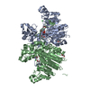 7qowC 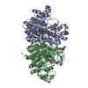 7qp8C 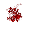 7z00C 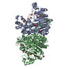 3e2dS S: Starting model for refinement C: citing same article ( |
|---|---|
| Similar structure data | |
| Experimental dataset #1 | Data reference: 10.5281/zenodo.5878920 / Data set type: diffraction image data |
-Links
 PDBj
PDBj
- Assembly
| Deposited unit | 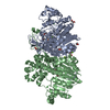
| ||||||||||||
|---|---|---|---|---|---|---|---|---|---|---|---|---|---|
| 1 |
| ||||||||||||
| Unit cell |
|
-Components
-Protein , 1 types, 2 molecules AB
| #1: Protein | Mass: 56626.094 Da / Num. of mol.: 2 Source method: isolated from a genetically manipulated source Details: Submitted sequence contains the C-terminal StrepTag-II included in the crystallised construct, but was not built into the model due to lack of electron density Source: (gene. exp.) Vibrio sp. G15-21 (bacteria) / Production host: |
|---|
-Non-polymers , 6 types, 791 molecules 


| #2: Chemical | ChemComp-ZN /  Mass: 65.409 Da / Num. of mol.: 4 Mass: 65.409 Da / Num. of mol.: 4Source method: isolated from a genetically manipulated source Formula: Zn / Source: (gene. exp.) Vibrio sp. G15-21 (bacteria) / Production host: #3: Chemical | ChemComp-MG /  Mass: 24.305 Da / Num. of mol.: 5 / Source method: obtained synthetically / Formula: Mg Mass: 24.305 Da / Num. of mol.: 5 / Source method: obtained synthetically / Formula: Mg#4: Chemical | ChemComp-EDO /  Mass: 62.068 Da / Num. of mol.: 11 / Source method: obtained synthetically / Formula: C2H6O2 Mass: 62.068 Da / Num. of mol.: 11 / Source method: obtained synthetically / Formula: C2H6O2#5: Chemical |  Mass: 94.971 Da / Num. of mol.: 2 / Source method: obtained synthetically / Formula: PO4 / Feature type: SUBJECT OF INVESTIGATION Mass: 94.971 Da / Num. of mol.: 2 / Source method: obtained synthetically / Formula: PO4 / Feature type: SUBJECT OF INVESTIGATION#6: Chemical |  Mass: 35.453 Da / Num. of mol.: 2 / Source method: obtained synthetically / Formula: Cl / Feature type: SUBJECT OF INVESTIGATION Mass: 35.453 Da / Num. of mol.: 2 / Source method: obtained synthetically / Formula: Cl / Feature type: SUBJECT OF INVESTIGATION#7: Water | ChemComp-HOH / | Mass: 18.015 Da / Num. of mol.: 767 / Source method: isolated from a natural source / Formula: H2O |
|---|
-Details
| Has ligand of interest | Y |
|---|
-Experimental details
-Experiment
| Experiment | Method: X-RAY DIFFRACTION / Number of used crystals: 1 |
|---|
- Sample preparation
| Crystal | Density Matthews: 2.26 Å3/Da / Density % sol: 45.6 % |
|---|---|
| Crystal grow | Temperature: 293 K / Method: vapor diffusion, hanging drop / pH: 7 / Details: 26% PEG3350, 0.5 M NaCl, 1.0 M Tris pH 7.0 |
-Data collection
| Diffraction | Mean temperature: 100 K / Serial crystal experiment: N |
|---|---|
| Diffraction source | Source: SYNCHROTRON / Site: PETRA III, DESY  / Beamline: P11 / Wavelength: 1.033 Å / Beamline: P11 / Wavelength: 1.033 Å |
| Detector | Type: DECTRIS PILATUS 6M-F / Detector: PIXEL / Date: Apr 20, 2021 |
| Radiation | Protocol: SINGLE WAVELENGTH / Monochromatic (M) / Laue (L): M / Scattering type: x-ray |
| Radiation wavelength | Wavelength: 1.033 Å / Relative weight: 1 |
| Reflection | Resolution: 1.29→39.31 Å / Num. obs: 1574856 / % possible obs: 96.8 % / Redundancy: 6.45 % / Biso Wilson estimate: 16.87 Å2 / CC1/2: 0.999 / Rrim(I) all: 0.057 / Net I/σ(I): 15.32 |
| Reflection shell | Resolution: 1.29→1.32 Å / Num. unique obs: 53734 / CC1/2: 0.584 |
- Processing
| Software |
| |||||||||||||||||||||||||||||||||||||||||||||||||||||||||||||||||||||||||||||||||||||||||||||||||||||||||
|---|---|---|---|---|---|---|---|---|---|---|---|---|---|---|---|---|---|---|---|---|---|---|---|---|---|---|---|---|---|---|---|---|---|---|---|---|---|---|---|---|---|---|---|---|---|---|---|---|---|---|---|---|---|---|---|---|---|---|---|---|---|---|---|---|---|---|---|---|---|---|---|---|---|---|---|---|---|---|---|---|---|---|---|---|---|---|---|---|---|---|---|---|---|---|---|---|---|---|---|---|---|---|---|---|---|---|
| Refinement | Method to determine structure: MOLECULAR REPLACEMENT Starting model: 3E2D Resolution: 1.29→39.31 Å / SU ML: 0.1058 / Cross valid method: FREE R-VALUE / σ(F): 1.35 / Phase error: 16.1181 Stereochemistry target values: GeoStd + Monomer Library + CDL v1.2
| |||||||||||||||||||||||||||||||||||||||||||||||||||||||||||||||||||||||||||||||||||||||||||||||||||||||||
| Solvent computation | Shrinkage radii: 0.9 Å / VDW probe radii: 1.11 Å / Solvent model: FLAT BULK SOLVENT MODEL | |||||||||||||||||||||||||||||||||||||||||||||||||||||||||||||||||||||||||||||||||||||||||||||||||||||||||
| Displacement parameters | Biso mean: 24.7 Å2 | |||||||||||||||||||||||||||||||||||||||||||||||||||||||||||||||||||||||||||||||||||||||||||||||||||||||||
| Refinement step | Cycle: LAST / Resolution: 1.29→39.31 Å
| |||||||||||||||||||||||||||||||||||||||||||||||||||||||||||||||||||||||||||||||||||||||||||||||||||||||||
| Refine LS restraints |
| |||||||||||||||||||||||||||||||||||||||||||||||||||||||||||||||||||||||||||||||||||||||||||||||||||||||||
| LS refinement shell |
|
