 Movie
Movie Controller

[English] 日本語
 Yorodumi
- PDB-7yzf: Crystal structure of the human FoxA2 bound to the TGTTTATT site (... -
+ Open data

- Basic information
| Entry | Database: PDB / ID: 7yzf | |||||||||
|---|---|---|---|---|---|---|---|---|---|---|
| Title | Crystal structure of the human FoxA2 bound to the TGTTTATT site (forkhead motif ATAAACA) | |||||||||
 Components Components |
| |||||||||
 Keywords Keywords | DNA BINDING PROTEIN / transcription factor | |||||||||
| Function / homology |  Function and homology information Function and homology informationPositive Regulation of CDH1 Gene Transcription / primitive streak formation / positive regulation of cell-cell adhesion mediated by cadherin / response to interleukin-6 / Developmental Lineage of Pancreatic Acinar Cells / positive regulation of embryonic development / Developmental Lineage of Multipotent Pancreatic Progenitor Cells / endocrine pancreas development / regulation of insulin secretion involved in cellular response to glucose stimulus / Formation of definitive endoderm ...Positive Regulation of CDH1 Gene Transcription / primitive streak formation / positive regulation of cell-cell adhesion mediated by cadherin / response to interleukin-6 / Developmental Lineage of Pancreatic Acinar Cells / positive regulation of embryonic development / Developmental Lineage of Multipotent Pancreatic Progenitor Cells / endocrine pancreas development / regulation of insulin secretion involved in cellular response to glucose stimulus / Formation of definitive endoderm / Formation of axial mesoderm / dopaminergic neuron differentiation / cell fate specification / positive regulation of gastrulation / negative regulation of epithelial to mesenchymal transition / regulation of blood coagulation / Regulation of gene expression in beta cells / anatomical structure morphogenesis / Developmental Lineage of Pancreatic Ductal Cells / adult locomotory behavior / DNA-binding transcription repressor activity, RNA polymerase II-specific / sequence-specific double-stranded DNA binding / transcription corepressor activity / chromatin organization / DNA-binding transcription activator activity, RNA polymerase II-specific / nucleic acid binding / RNA polymerase II-specific DNA-binding transcription factor binding / DNA-binding transcription factor activity, RNA polymerase II-specific / cell differentiation / transcription cis-regulatory region binding / RNA polymerase II cis-regulatory region sequence-specific DNA binding / DNA-binding transcription factor activity / protein domain specific binding / regulation of transcription by RNA polymerase II / positive regulation of DNA-templated transcription / chromatin / DNA-templated transcription / negative regulation of transcription by RNA polymerase II / positive regulation of transcription by RNA polymerase II / DNA binding / nucleoplasm / nucleus / cytoplasm Similarity search - Function | |||||||||
| Biological species |  Homo sapiens (human) Homo sapiens (human) | |||||||||
| Method |  X-RAY DIFFRACTION / SYNCHROTRON / MOLECULAR REPLACEMENT / Resolution: 2.18 Å X-RAY DIFFRACTION / SYNCHROTRON / MOLECULAR REPLACEMENT / Resolution: 2.18 Å | |||||||||
 Authors Authors | Pluta, R. / Macias, M.J. | |||||||||
| Funding support |  Spain, European Union, 2items Spain, European Union, 2items
| |||||||||
 Citation Citation | Journal: Nat Commun / Year: 2022 Title: Molecular basis for DNA recognition by the maternal pioneer transcription factor FoxH1. Authors: Pluta, R. / Aragon, E. / Prescott, N.A. / Ruiz, L. / Mees, R.A. / Baginski, B. / Flood, J.R. / Martin-Malpartida, P. / Massague, J. / David, Y. / Macias, M.J. | |||||||||
| History |
|
- Structure visualization
| Structure viewer | Molecule: MolmilJmol/JSmol |
|---|
- Downloads & links
-Download
| PDBx/mmCIF format | 7yzf.cif.gz | 89.5 KB | Display | PDBx/mmCIF format |
|---|---|---|---|---|
| PDB format | pdb7yzf.ent.gz | 63.7 KB | Display | PDB format |
| PDBx/mmJSON format | 7yzf.json.gz | Tree view | PDBx/mmJSON format | |
| Others |  Other downloads Other downloads |
-Validation report
| Arichive directory | https://data.pdbj.org/pub/pdb/validation_reports/yz/7yzfftp://data.pdbj.org/pub/pdb/validation_reports/yz/7yzf | HTTPS FTP |
|---|
-Related structure data
| Related structure data |  7yz7C  7yzaC  7yzbC  7yzcC  7yzdC  7yzeSC  7yzgC S: Starting model for refinement C: citing same article ( |
|---|---|
| Similar structure data |
-Links
 PDBj
PDBj


- Assembly
| Deposited unit | 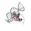
| ||||||||
|---|---|---|---|---|---|---|---|---|---|
| 1 |
| ||||||||
| Unit cell |
|
-Components
| #1: DNA chain | Mass: 4967.250 Da / Num. of mol.: 1 / Source method: obtained synthetically / Source: (synth.) Homo sapiens (human) |
|---|---|
| #2: DNA chain | Mass: 4825.181 Da / Num. of mol.: 1 / Source method: obtained synthetically / Source: (synth.) Homo sapiens (human) |
| #3: Protein | Mass: 14652.791 Da / Num. of mol.: 1 Source method: isolated from a genetically manipulated source Source: (gene. exp.) Homo sapiens (human) / Gene: FOXA2, HNF3B, TCF3B / Production host:  |
| #4: Chemical | ChemComp-K /   Mass: 39.098 Da / Num. of mol.: 1 / Source method: obtained synthetically / Formula: K / Feature type: SUBJECT OF INVESTIGATION Mass: 39.098 Da / Num. of mol.: 1 / Source method: obtained synthetically / Formula: K / Feature type: SUBJECT OF INVESTIGATION |
| #5: Water | ChemComp-HOH /  Mass: 18.015 Da / Num. of mol.: 5 / Source method: isolated from a natural source / Formula: H2O Mass: 18.015 Da / Num. of mol.: 5 / Source method: isolated from a natural source / Formula: H2O |
| Has ligand of interest | Y |
-Experimental details
-Experiment
| Experiment | Method: X-RAY DIFFRACTION / Number of used crystals: 1 |
|---|
- Sample preparation
| Crystal | Density Matthews: 2.52 Å3/Da / Density % sol: 51.14 % |
|---|---|
| Crystal grow | Temperature: 277 K / Method: vapor diffusion, sitting drop / pH: 5.5 / Details: 25% PEG 3350, 0.1 M Bis-Tris pH 5.5 |
-Data collection
| Diffraction | Mean temperature: 100 K / Serial crystal experiment: N |
|---|---|
| Diffraction source | Source: SYNCHROTRON / Site: ALBA / Beamline: XALOC / Wavelength: 0.97926 Å |
| Detector | Type: DECTRIS PILATUS 6M / Detector: PIXEL / Date: Feb 1, 2020 |
| Radiation | Protocol: SINGLE WAVELENGTH / Monochromatic (M) / Laue (L): M / Scattering type: x-ray |
| Radiation wavelength | Wavelength: 0.97926 Å / Relative weight: 1 |
| Reflection | Resolution: 2.179→58.421 Å / Num. obs: 9215 / % possible obs: 86.5 % / Redundancy: 9.9 % / CC1/2: 0.998 / Rpim(I) all: 0.018 / Net I/σ(I): 19.9 |
| Reflection shell | Resolution: 2.179→2.378 Å / Num. unique obs: 462 / CC1/2: 0.593 / Rpim(I) all: 0.544 / % possible all: 52.2 |
- Processing
| Software |
| |||||||||||||||||||||||||||||||||||||||||||||||||||||||||||||||||||||||||||||||||||||||||||||||||||||||||
|---|---|---|---|---|---|---|---|---|---|---|---|---|---|---|---|---|---|---|---|---|---|---|---|---|---|---|---|---|---|---|---|---|---|---|---|---|---|---|---|---|---|---|---|---|---|---|---|---|---|---|---|---|---|---|---|---|---|---|---|---|---|---|---|---|---|---|---|---|---|---|---|---|---|---|---|---|---|---|---|---|---|---|---|---|---|---|---|---|---|---|---|---|---|---|---|---|---|---|---|---|---|---|---|---|---|---|
| Refinement | Method to determine structure: MOLECULAR REPLACEMENT Starting model: 7YZE Resolution: 2.18→58.42 Å / Cor.coef. Fo:Fc: 0.948 / Cor.coef. Fo:Fc free: 0.933 / SU B: 18.039 / SU ML: 0.194 / Cross valid method: THROUGHOUT / ESU R: 0.401 / ESU R Free: 0.268 / Stereochemistry target values: MAXIMUM LIKELIHOOD Details: U VALUES : WITH TLS ADDED HYDROGENS HAVE BEEN ADDED IN THE RIDING POSITIONS U VALUES : RESIDUAL ONLY
| |||||||||||||||||||||||||||||||||||||||||||||||||||||||||||||||||||||||||||||||||||||||||||||||||||||||||
| Solvent computation | Ion probe radii: 0.7 Å / Shrinkage radii: 0.7 Å / VDW probe radii: 1 Å / Solvent model: MASK | |||||||||||||||||||||||||||||||||||||||||||||||||||||||||||||||||||||||||||||||||||||||||||||||||||||||||
| Displacement parameters | Biso mean: 72.04 Å2
| |||||||||||||||||||||||||||||||||||||||||||||||||||||||||||||||||||||||||||||||||||||||||||||||||||||||||
| Refinement step | Cycle: LAST / Resolution: 2.18→58.42 Å
| |||||||||||||||||||||||||||||||||||||||||||||||||||||||||||||||||||||||||||||||||||||||||||||||||||||||||
| Refine LS restraints |
| |||||||||||||||||||||||||||||||||||||||||||||||||||||||||||||||||||||||||||||||||||||||||||||||||||||||||
| LS refinement shell | Resolution: 2.18→2.236 Å
| |||||||||||||||||||||||||||||||||||||||||||||||||||||||||||||||||||||||||||||||||||||||||||||||||||||||||
| Refinement TLS params. | Method: refined / Refine-ID: X-RAY DIFFRACTION
| |||||||||||||||||||||||||||||||||||||||||||||||||||||||||||||||||||||||||||||||||||||||||||||||||||||||||
| Refinement TLS group |
|
