 Movie
Movie Controller

[English] 日本語
 Yorodumi
- PDB-7yy8: Crystal structure of Mycobacterium abscessus Phosphopantetheine a... -
+ Open data

- Basic information
| Entry | Database: PDB / ID: 7yy8 | ||||||
|---|---|---|---|---|---|---|---|
| Title | Crystal structure of Mycobacterium abscessus Phosphopantetheine adenylyltransferase in complex with Fragment 12 | ||||||
 Components Components | Phosphopantetheine adenylyltransferase | ||||||
 Keywords Keywords | TRANSFERASE / CoaD / PPAT / nucleotidyltransferase | ||||||
| Function / homology |  Function and homology information Function and homology informationpantetheine-phosphate adenylyltransferase / pantetheine-phosphate adenylyltransferase activity / coenzyme A biosynthetic process / ATP binding / cytoplasm Similarity search - Function | ||||||
| Biological species |  Mycobacteroides abscessus (bacteria) Mycobacteroides abscessus (bacteria) | ||||||
| Method |  X-RAY DIFFRACTION / SYNCHROTRON / MOLECULAR REPLACEMENT / molecular replacement / Resolution: 1.516 Å X-RAY DIFFRACTION / SYNCHROTRON / MOLECULAR REPLACEMENT / molecular replacement / Resolution: 1.516 Å | ||||||
 Authors Authors | Thomas, S.E. / Coyne, A.G. / Blundell, T.L. | ||||||
| Funding support |  United Kingdom, 1items United Kingdom, 1items
| ||||||
 Citation Citation | Journal: Front Mol Biosci / Year: 2022 Title: Structural Characterization of Mycobacterium abscessus Phosphopantetheine Adenylyl Transferase Ligand Interactions: Implications for Fragment-Based Drug Design. Authors: Thomas, S.E. / McCarthy, W.J. / El Bakali, J. / Brown, K.P. / Kim, S.Y. / Blaszczyk, M. / Mendes, V. / Abell, C. / Floto, R.A. / Coyne, A.G. / Blundell, T.L. | ||||||
| History |
|
- Structure visualization
| Structure viewer | Molecule: MolmilJmol/JSmol |
|---|
- Downloads & links
-Download
| PDBx/mmCIF format | 7yy8.cif.gz | 110.4 KB | Display | PDBx/mmCIF format |
|---|---|---|---|---|
| PDB format | pdb7yy8.ent.gz | 83.8 KB | Display | PDB format |
| PDBx/mmJSON format | 7yy8.json.gz | Tree view | PDBx/mmJSON format | |
| Others |  Other downloads Other downloads |
-Validation report
| Arichive directory | https://data.pdbj.org/pub/pdb/validation_reports/yy/7yy8ftp://data.pdbj.org/pub/pdb/validation_reports/yy/7yy8 | HTTPS FTP |
|---|
-Related structure data
| Related structure data | 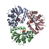 7ywmC  7yxzC 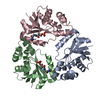 7yy0C 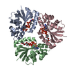 7yy1C 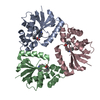 7yy2C  7yy3C 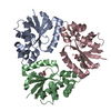 7yy4C  7yy5C  7yy6C 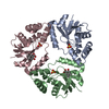 7yy7C 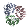 7yy9C  7yyaC 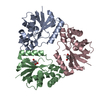 7yybC  7yycC  5o06S S: Starting model for refinement C: citing same article ( |
|---|---|
| Similar structure data |
-Links
 PDBj
PDBj- Assembly
| Deposited unit | 
| |||||||||
|---|---|---|---|---|---|---|---|---|---|---|
| 1 | 
| |||||||||
| Unit cell |
| |||||||||
| Components on special symmetry positions |
|
-Components
| #1: Protein | Mass: 17476.887 Da / Num. of mol.: 3 Source method: isolated from a genetically manipulated source Source: (gene. exp.) Mycobacteroides abscessus (bacteria) / Gene: coaD, MAB_3259c / Production host: References: UniProt: B1MDL6, pantetheine-phosphate adenylyltransferase #2: Chemical | ChemComp-3IL / | 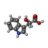  Mass: 205.210 Da / Num. of mol.: 1 / Source method: obtained synthetically / Formula: C11H11NO3 / Feature type: SUBJECT OF INVESTIGATION Mass: 205.210 Da / Num. of mol.: 1 / Source method: obtained synthetically / Formula: C11H11NO3 / Feature type: SUBJECT OF INVESTIGATION#3: Chemical | ChemComp-4Z9 / ( | 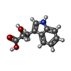  Mass: 205.210 Da / Num. of mol.: 1 / Source method: obtained synthetically / Formula: C11H11NO3 Mass: 205.210 Da / Num. of mol.: 1 / Source method: obtained synthetically / Formula: C11H11NO3#4: Water | ChemComp-HOH / |  Mass: 18.015 Da / Num. of mol.: 338 / Source method: isolated from a natural source / Formula: H2O Mass: 18.015 Da / Num. of mol.: 338 / Source method: isolated from a natural source / Formula: H2OHas ligand of interest | Y | |
|---|
-Experimental details
-Experiment
| Experiment | Method: X-RAY DIFFRACTION / Number of used crystals: 1 |
|---|
- Sample preparation
| Crystal | Density Matthews: 2.7 Å3/Da / Density % sol: 54.37 % |
|---|---|
| Crystal grow | Temperature: 292 K / Method: vapor diffusion, sitting drop Details: 0.2 M Sodium bromide, 20-24 % PEG3350, 0.1 M Bis-Tris propane pH 6.5 |
-Data collection
| Diffraction | Mean temperature: 100 K / Serial crystal experiment: N | ||||||||||||||||||||||||||||||
|---|---|---|---|---|---|---|---|---|---|---|---|---|---|---|---|---|---|---|---|---|---|---|---|---|---|---|---|---|---|---|---|
| Diffraction source | Source: SYNCHROTRON / Site: Diamond / Beamline: I24 / Wavelength: 0.9686 Å | ||||||||||||||||||||||||||||||
| Detector | Type: DECTRIS PILATUS3 6M / Detector: PIXEL / Date: Aug 10, 2016 | ||||||||||||||||||||||||||||||
| Radiation | Protocol: SINGLE WAVELENGTH / Monochromatic (M) / Laue (L): M / Scattering type: x-ray | ||||||||||||||||||||||||||||||
| Radiation wavelength | Wavelength: 0.9686 Å / Relative weight: 1 | ||||||||||||||||||||||||||||||
| Reflection | Resolution: 1.516→119.16 Å / Num. obs: 87328 / % possible obs: 99.6 % / Redundancy: 7.8 % / CC1/2: 0.996 / Rmerge(I) obs: 0.102 / Rpim(I) all: 0.039 / Rrim(I) all: 0.109 / Net I/σ(I): 11.9 | ||||||||||||||||||||||||||||||
| Reflection shell | Diffraction-ID: 1
|
-Phasing
| Phasing | Method: molecular replacement |
|---|---|
| Phasing MR | Model details: Phaser MODE: MR_AUTO |
- Processing
| Software |
| ||||||||||||||||||||||||||||||||||||||||||||||||||||||||||||||||||||||||||||||||||||||||||||||||||||||||||||||||||||||||||||||||||||||||||||||||||||||||||||||||||||||||||||||||||||||||||
|---|---|---|---|---|---|---|---|---|---|---|---|---|---|---|---|---|---|---|---|---|---|---|---|---|---|---|---|---|---|---|---|---|---|---|---|---|---|---|---|---|---|---|---|---|---|---|---|---|---|---|---|---|---|---|---|---|---|---|---|---|---|---|---|---|---|---|---|---|---|---|---|---|---|---|---|---|---|---|---|---|---|---|---|---|---|---|---|---|---|---|---|---|---|---|---|---|---|---|---|---|---|---|---|---|---|---|---|---|---|---|---|---|---|---|---|---|---|---|---|---|---|---|---|---|---|---|---|---|---|---|---|---|---|---|---|---|---|---|---|---|---|---|---|---|---|---|---|---|---|---|---|---|---|---|---|---|---|---|---|---|---|---|---|---|---|---|---|---|---|---|---|---|---|---|---|---|---|---|---|---|---|---|---|---|---|---|---|
| Refinement | Method to determine structure: MOLECULAR REPLACEMENT Starting model: 5O06 Resolution: 1.516→56.979 Å / SU ML: 0.19 / Cross valid method: THROUGHOUT / σ(F): 1.35 / Phase error: 24.66 / Stereochemistry target values: ML
| ||||||||||||||||||||||||||||||||||||||||||||||||||||||||||||||||||||||||||||||||||||||||||||||||||||||||||||||||||||||||||||||||||||||||||||||||||||||||||||||||||||||||||||||||||||||||||
| Solvent computation | Shrinkage radii: 0.9 Å / VDW probe radii: 1.11 Å / Solvent model: FLAT BULK SOLVENT MODEL | ||||||||||||||||||||||||||||||||||||||||||||||||||||||||||||||||||||||||||||||||||||||||||||||||||||||||||||||||||||||||||||||||||||||||||||||||||||||||||||||||||||||||||||||||||||||||||
| Displacement parameters | Biso max: 84.67 Å2 / Biso mean: 28.1458 Å2 / Biso min: 12.63 Å2 | ||||||||||||||||||||||||||||||||||||||||||||||||||||||||||||||||||||||||||||||||||||||||||||||||||||||||||||||||||||||||||||||||||||||||||||||||||||||||||||||||||||||||||||||||||||||||||
| Refinement step | Cycle: final / Resolution: 1.516→56.979 Å
| ||||||||||||||||||||||||||||||||||||||||||||||||||||||||||||||||||||||||||||||||||||||||||||||||||||||||||||||||||||||||||||||||||||||||||||||||||||||||||||||||||||||||||||||||||||||||||
| Refine LS restraints |
| ||||||||||||||||||||||||||||||||||||||||||||||||||||||||||||||||||||||||||||||||||||||||||||||||||||||||||||||||||||||||||||||||||||||||||||||||||||||||||||||||||||||||||||||||||||||||||
| LS refinement shell | Refine-ID: X-RAY DIFFRACTION / Rfactor Rfree error: 0
|
