Entry Database : PDB / ID : 7yxwTitle Structure of the p22phox A200G mutant in complex with p47phox peptide Cytochrome b-245 light chain Neutrophil cytosol factor 1 Keywords / / Function / homology Function Domain/homology Component
/ / / / / / / / / / / / / / / / / / / / / / / / / / / / / / / / / / / / / / / / / / / / / / / / / / / / / / / / / / / / / / / / / / / / / / / / / / / / / / / / / / / / / / / / / / / / / / / / / / / / / / / / / / / / / / / / / / / / / / / / / Biological species Homo sapiens (human)Method / / / Resolution : 2.5 Å Authors Cukier, C.D. / Vuillard, L.M. / Komjati, B. / Szlavik, Z. Funding support 1items Organization Grant number Country Not funded
Journal : Acs Med.Chem.Lett. / Year : 2022Title : Targeting NOX2 via p47/phox-p22/phox Inhibition with Novel Triproline MimeticsAuthors: Garsi, J.B. / Komjati, B. / Cullia, G. / Fejes, I. / Sipos, M. / Sipos, Z. / Fordos, E. / Markacz, P. / Balazs, B. / Lancelot, N. / Berger, S. / Raimbaud, E. / Brown, D. / Vuillard, L.M. / ... Authors : Garsi, J.B. / Komjati, B. / Cullia, G. / Fejes, I. / Sipos, M. / Sipos, Z. / Fordos, E. / Markacz, P. / Balazs, B. / Lancelot, N. / Berger, S. / Raimbaud, E. / Brown, D. / Vuillard, L.M. / Haberkorn, L. / Cukier, C. / Szlavik, Z. / Hanessian, S. History Deposition Feb 16, 2022 Deposition site / Processing site Revision 1.0 Mar 30, 2022 Provider / Type Revision 1.1 Jun 22, 2022 Group / Category / citation_authorItem _citation.country / _citation.journal_abbrev ... _citation.country / _citation.journal_abbrev / _citation.journal_id_CSD / _citation.journal_id_ISSN / _citation.journal_volume / _citation.page_first / _citation.page_last / _citation.pdbx_database_id_DOI / _citation.title / _citation.year Revision 1.2 Jan 31, 2024 Group / Refinement descriptionCategory / chem_comp_bond / pdbx_initial_refinement_model
Show all Show less
 Movie
Movie Controller

 Yorodumi
 Open data
 Basic information
 Components
 Keywords
 Function and homology information
 Homo sapiens (human)
 X-RAY DIFFRACTION /
  Authors
 Citation
 Structure visualization
 Downloads & links
 Other downloads
 PDBj
PDBj


 Assembly
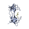

 Sample preparation
 / Beamline: PROXIMA 1 / Wavelength: 0.9786 Å
 Processing