 Movie
Movie Controller


+ Open data

- Basic information
| Entry | Database: PDB / ID: 7ywn | |||||||||||||||
|---|---|---|---|---|---|---|---|---|---|---|---|---|---|---|---|---|
| Title | Six DNA Helix Bundle nanopore - State 4 | |||||||||||||||
 Components Components | (DNA (50-MER)) x 6 | |||||||||||||||
 Keywords Keywords | DNA / DNA origami / nanopore. | |||||||||||||||
| Function / homology | DNA / DNA (> 10) Function and homology information Function and homology information | |||||||||||||||
| Biological species | DNA molecule (others) | |||||||||||||||
| Method | ELECTRON MICROSCOPY / single particle reconstruction / cryo EM / Resolution: 8 Å | |||||||||||||||
 Authors Authors | Javed, A. / Ahmad, K. / Lanphere, C. / Orlova, E.V. / Coveney, P. / Howorka, S. | |||||||||||||||
| Funding support |  United Kingdom, 4items United Kingdom, 4items
| |||||||||||||||
 Citation Citation | Journal: Nat Commun / Year: 2023 Title: Structure and dynamics of an archetypal DNA nanoarchitecture revealed via cryo-EM and molecular dynamics simulations. Authors: Katya Ahmad / Abid Javed / Conor Lanphere / Peter V Coveney / Elena V Orlova / Stefan Howorka /  Abstract: DNA can be folded into rationally designed, unique, and functional materials. To fully realise the potential of these DNA materials, a fundamental understanding of their structure and dynamics is ...DNA can be folded into rationally designed, unique, and functional materials. To fully realise the potential of these DNA materials, a fundamental understanding of their structure and dynamics is necessary, both in simple solvents as well as more complex and diverse anisotropic environments. Here we analyse an archetypal six-duplex DNA nanoarchitecture with single-particle cryo-electron microscopy and molecular dynamics simulations in solvents of tunable ionic strength and within the anisotropic environment of biological membranes. Outside lipid bilayers, the six-duplex bundle lacks the designed symmetrical barrel-type architecture. Rather, duplexes are arranged in non-hexagonal fashion and are disorted to form a wider, less elongated structure. Insertion into lipid membranes, however, restores the anticipated barrel shape due to lateral duplex compression by the bilayer. The salt concentration has a drastic impact on the stability of the inserted barrel-shaped DNA nanopore given the tunable electrostatic repulsion between the negatively charged duplexes. By synergistically combining experiments and simulations, we increase fundamental understanding into the environment-dependent structural dynamics of a widely used nanoarchitecture. This insight will pave the way for future engineering and biosensing applications. | |||||||||||||||
| History |
|
- Structure visualization
| Structure viewer | Molecule: MolmilJmol/JSmol |
|---|
- Downloads & links
-Download
| PDBx/mmCIF format | 7ywn.cif.gz | 129.8 KB | Display | PDBx/mmCIF format |
|---|---|---|---|---|
| PDB format | pdb7ywn.ent.gz | 101.8 KB | Display | PDB format |
| PDBx/mmJSON format | 7ywn.json.gz | Tree view | PDBx/mmJSON format | |
| Others |  Other downloads Other downloads |
-Validation report
| Arichive directory | https://data.pdbj.org/pub/pdb/validation_reports/yw/7ywnftp://data.pdbj.org/pub/pdb/validation_reports/yw/7ywn | HTTPS FTP |
|---|
-Related structure data
| Related structure data |  14345MC  7ywhC 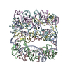 7ywiC  7ywlC  7ywoC C: citing same article ( M: map data used to model this data |
|---|---|
| Similar structure data |


-Links
 PDBj
PDBj


- Assembly
| Deposited unit | 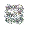
|
|---|---|
| 1 |
|
-Components
-DNA chain , 6 types, 6 molecules ABCDEF
| #1: DNA chain | Mass: 15334.821 Da / Num. of mol.: 1 / Source method: obtained synthetically / Source: (synth.) DNA molecule (others) |
|---|---|
| #2: DNA chain | Mass: 15413.827 Da / Num. of mol.: 1 / Source method: obtained synthetically / Source: (synth.) DNA molecule (others) |
| #3: DNA chain | Mass: 15285.784 Da / Num. of mol.: 1 / Source method: obtained synthetically / Source: (synth.) DNA molecule (others) |
| #4: DNA chain | Mass: 15483.923 Da / Num. of mol.: 1 / Source method: obtained synthetically / Source: (synth.) DNA molecule (others) |
| #5: DNA chain | Mass: 15403.877 Da / Num. of mol.: 1 / Source method: obtained synthetically / Source: (synth.) DNA molecule (others) |
| #6: DNA chain | Mass: 15268.795 Da / Num. of mol.: 1 / Source method: obtained synthetically / Source: (synth.) DNA molecule (others) |
-Experimental details
-Experiment
| Experiment | Method: ELECTRON MICROSCOPY |
|---|---|
| EM experiment | Aggregation state: PARTICLE / 3D reconstruction method: single particle reconstruction |
- Sample preparation
| Component | Name: Six DNA helix bundle DNA nanopore / Type: COMPLEX / Entity ID: all / Source: MULTIPLE SOURCES |
|---|---|
| Buffer solution | pH: 7.4 |
| Buffer component | Conc.: 12 mM / Name: Magnesium Chloride / Formula: MgCl2 |
| Specimen | Conc.: 1 mg/ml / Embedding applied: NO / Shadowing applied: NO / Staining applied: NO / Vitrification applied: YES |
| Specimen support | Grid material: COPPER / Grid mesh size: 300 divisions/in. / Grid type: C-flat-2/2 |
| Vitrification | Instrument: FEI VITROBOT MARK IV / Cryogen name: ETHANE / Humidity: 100 % / Chamber temperature: 283 K |
- Electron microscopy imaging
| Experimental equipment |  Model: Titan Krios / Image courtesy: FEI Company |
|---|---|
| Microscopy | Model: FEI TITAN KRIOS |
| Electron gun | Electron source:  FIELD EMISSION GUN / Accelerating voltage: 300 kV / Illumination mode: FLOOD BEAM FIELD EMISSION GUN / Accelerating voltage: 300 kV / Illumination mode: FLOOD BEAM |
| Electron lens | Mode: BRIGHT FIELD / Calibrated magnification: 47000 X / Nominal defocus max: 2500 nm / Nominal defocus min: 1200 nm / C2 aperture diameter: 70 µm / Alignment procedure: ZEMLIN TABLEAU |
| Specimen holder | Cryogen: NITROGEN / Specimen holder model: FEI TITAN KRIOS AUTOGRID HOLDER |
| Image recording | Electron dose: 1.2 e/Å2 / Detector mode: COUNTING / Film or detector model: GATAN K2 SUMMIT (4k x 4k) / Num. of grids imaged: 1 |
| Image scans | Sampling size: 5 µm / Movie frames/image: 50 |
- Processing
| EM software |
| ||||||||||||||||||||||||||||||||||||||||||||
|---|---|---|---|---|---|---|---|---|---|---|---|---|---|---|---|---|---|---|---|---|---|---|---|---|---|---|---|---|---|---|---|---|---|---|---|---|---|---|---|---|---|---|---|---|---|
| CTF correction | Type: PHASE FLIPPING AND AMPLITUDE CORRECTION | ||||||||||||||||||||||||||||||||||||||||||||
| Particle selection | Num. of particles selected: 368501 | ||||||||||||||||||||||||||||||||||||||||||||
| Symmetry | Point symmetry: C1 (asymmetric) | ||||||||||||||||||||||||||||||||||||||||||||
| 3D reconstruction | Resolution: 8 Å / Resolution method: FSC 0.5 CUT-OFF / Num. of particles: 27900 / Algorithm: FOURIER SPACE / Num. of class averages: 1 / Symmetry type: POINT | ||||||||||||||||||||||||||||||||||||||||||||
| Atomic model building | Protocol: FLEXIBLE FIT / Space: REAL / Target criteria: Cross-correlation |
