 Movie
Movie Controller


+ Open data

- Basic information
| Entry | Database: PDB / ID: 7ypx | ||||||
|---|---|---|---|---|---|---|---|
| Title | Cyanophage Pam3 fiber | ||||||
 Components Components |
| ||||||
 Keywords Keywords | VIRAL PROTEIN / fiber / VIRUS | ||||||
| Biological species |  uncultured cyanophage (environmental samples) uncultured cyanophage (environmental samples) | ||||||
| Method | ELECTRON MICROSCOPY / single particle reconstruction / cryo EM / Resolution: 3.12 Å | ||||||
 Authors Authors | Wei, Z.L. / Jiang, Y.L. / Zhou, C.Z. | ||||||
| Funding support |  China, 1items China, 1items
| ||||||
 Citation Citation | Journal: Viruses / Year: 2022 Title: Structural Insights into the Chaperone-Assisted Assembly of a Simplified Tail Fiber of the Myocyanophage Pam3. Authors: Zi-Lu Wei / Feng Yang / Bo Li / Pu Hou / Wen-Wen Kong / Jie Wang / Yuxing Chen / Yong-Liang Jiang / Cong-Zhao Zhou / Abstract: At the first step of phage infection, the receptor-binding proteins (RBPs) such as tail fibers are responsible for recognizing specific host surface receptors. The proper folding and assembly of tail ...At the first step of phage infection, the receptor-binding proteins (RBPs) such as tail fibers are responsible for recognizing specific host surface receptors. The proper folding and assembly of tail fibers usually requires a chaperone encoded by the phage genome. Despite extensive studies on phage structures, the molecular mechanism of phage tail fiber assembly remains largely unknown. Here, using a minimal myocyanophage, termed Pam3, isolated from Lake Chaohu, we demonstrate that the chaperone gp25 forms a stable complex with the tail fiber gp24 at a stoichiometry of 3:3. The 3.1-Å cryo-electron microscopy structure of this complex revealed an elongated structure with the gp25 trimer embracing the distal moieties of gp24 trimer at the center. Each gp24 subunit consists of three domains: the N-terminal α-helical domain required for docking to the baseplate, the tumor necrosis factor (TNF)-like and glycine-rich domains responsible for recognizing the host receptor. Each gp25 subunit consists of two domains: a non-conserved N-terminal β-sandwich domain that binds to the TNF-like and glycine-rich domains of the fiber, and a C-terminal α-helical domain that mediates trimerization/assembly of the fiber. Structural analysis enabled us to propose the assembly mechanism of phage tail fibers, in which the chaperone first protects the intertwined and repetitive distal moiety of each fiber subunit, further ensures the proper folding of these highly plastic structural elements, and eventually enables the formation of the trimeric fiber. These findings provide the structural basis for the design and engineering of phage fibers for biotechnological applications. | ||||||
| History |
|
- Structure visualization
| Structure viewer | Molecule:  MolmilJmol/JSmol MolmilJmol/JSmol |
|---|
- Downloads & links
-Download
| PDBx/mmCIF format | 7ypx.cif.gz | 198.8 KB | Display | PDBx/mmCIF format |
|---|---|---|---|---|
| PDB format | pdb7ypx.ent.gz | 161.3 KB | Display | PDB format |
| PDBx/mmJSON format | 7ypx.json.gz | Tree view | PDBx/mmJSON format | |
| Others |  Other downloads Other downloads |
-Validation report
| Arichive directory | https://data.pdbj.org/pub/pdb/validation_reports/yp/7ypxftp://data.pdbj.org/pub/pdb/validation_reports/yp/7ypx | HTTPS FTP |
|---|
-Related structure data
-Links
 PDBj
PDBj- Assembly
| Deposited unit | 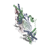
|
|---|---|
| 1 |
|
-Components
| #1: Protein | Mass: 28383.873 Da / Num. of mol.: 3 Source method: isolated from a genetically manipulated source Details: The source organism is the myocyanophage Pam3 isolated from the Lake Chaohu, China. Source: (gene. exp.) uncultured cyanophage (environmental samples)Production host:  #2: Protein | Mass: 17457.660 Da / Num. of mol.: 3 Source method: isolated from a genetically manipulated source Details: The source organism is the myocyanophage Pam3 isolated from the Lake Chaohu, China. Source: (gene. exp.) uncultured cyanophage (environmental samples)Production host: |
|---|
-Experimental details
-Experiment
| Experiment | Method: ELECTRON MICROSCOPY |
|---|---|
| EM experiment | Aggregation state: PARTICLE / 3D reconstruction method: single particle reconstruction |
- Sample preparation
| Component | Name: Pam3 tail fiber / Type: COMPLEX / Entity ID: all / Source: RECOMBINANT |
|---|---|
| Source (natural) | Organism: uncultured cyanophage (environmental samples) |
| Source (recombinant) | Organism: |
| Buffer solution | pH: 7 |
| Specimen | Embedding applied: NO / Shadowing applied: NO / Staining applied: NO / Vitrification applied: YES |
| Vitrification | Cryogen name: ETHANE / Humidity: 100 % / Chamber temperature: 300 K |
- Electron microscopy imaging
| Experimental equipment |  Model: Titan Krios / Image courtesy: FEI Company |
|---|---|
| Microscopy | Model: FEI TITAN KRIOS |
| Electron gun | Electron source:  FIELD EMISSION GUN / Accelerating voltage: 300 kV / Illumination mode: SPOT SCAN FIELD EMISSION GUN / Accelerating voltage: 300 kV / Illumination mode: SPOT SCAN |
| Electron lens | Mode: BRIGHT FIELD / Nominal defocus max: 2500 nm / Nominal defocus min: 1500 nm |
| Image recording | Electron dose: 55 e/Å2 / Film or detector model: GATAN K3 (6k x 4k) |
- Processing
| Software | Name: PHENIX / Version: 1.19.2_4158: / Classification: refinement | ||||||||||||||||||||||||
|---|---|---|---|---|---|---|---|---|---|---|---|---|---|---|---|---|---|---|---|---|---|---|---|---|---|
| CTF correction | Type: PHASE FLIPPING ONLY | ||||||||||||||||||||||||
| 3D reconstruction | Resolution: 3.12 Å / Resolution method: FSC 0.143 CUT-OFF / Num. of particles: 243989 / Symmetry type: POINT | ||||||||||||||||||||||||
| Refine LS restraints |
|
