 Movie
Movie Controller


+ Open data

- Basic information
| Entry | Database: PDB / ID: 7ypg | ||||||
|---|---|---|---|---|---|---|---|
| Title | Cryo-EM structure of amyloid fibril formed by tau (297-391) | ||||||
 Components Components | Isoform Tau-E of Microtubule-associated protein tau | ||||||
 Keywords Keywords | PROTEIN FIBRIL / amyloid fibril | ||||||
| Function / homology |  Function and homology information Function and homology informationplus-end-directed organelle transport along microtubule / histone-dependent DNA binding / negative regulation of protein localization to mitochondrion / neurofibrillary tangle / microtubule lateral binding / axonal transport / tubulin complex / positive regulation of protein localization to synapse / phosphatidylinositol bisphosphate binding / generation of neurons ...plus-end-directed organelle transport along microtubule / histone-dependent DNA binding / negative regulation of protein localization to mitochondrion / neurofibrillary tangle / microtubule lateral binding / axonal transport / tubulin complex / positive regulation of protein localization to synapse / phosphatidylinositol bisphosphate binding / generation of neurons / rRNA metabolic process / axonal transport of mitochondrion / regulation of mitochondrial fission / axon development / regulation of microtubule-based movement / intracellular distribution of mitochondria / regulation of chromosome organization / central nervous system neuron development / minor groove of adenine-thymine-rich DNA binding / lipoprotein particle binding / microtubule polymerization / negative regulation of mitochondrial membrane potential / regulation of microtubule polymerization / dynactin binding / apolipoprotein binding / protein polymerization / main axon / Caspase-mediated cleavage of cytoskeletal proteins / regulation of microtubule polymerization or depolymerization / negative regulation of mitochondrial fission / axolemma / glial cell projection / neurofibrillary tangle assembly / positive regulation of axon extension / regulation of cellular response to heat / positive regulation of microtubule polymerization / positive regulation of protein localization / Activation of AMPK downstream of NMDARs / positive regulation of superoxide anion generation / cellular response to brain-derived neurotrophic factor stimulus / regulation of long-term synaptic depression / supramolecular fiber organization / cytoplasmic microtubule organization / regulation of calcium-mediated signaling / axon cytoplasm / somatodendritic compartment / synapse assembly / phosphatidylinositol binding / nuclear periphery / astrocyte activation / enzyme inhibitor activity / protein phosphatase 2A binding / stress granule assembly / regulation of microtubule cytoskeleton organization / regulation of autophagy / cellular response to reactive oxygen species / microglial cell activation / cellular response to nerve growth factor stimulus / Hsp90 protein binding / protein homooligomerization / SH3 domain binding / PKR-mediated signaling / synapse organization / regulation of synaptic plasticity / response to lead ion / microtubule cytoskeleton organization / memory / neuron projection development / cytoplasmic ribonucleoprotein granule / cell-cell signaling / single-stranded DNA binding / cellular response to heat / protein-folding chaperone binding / growth cone / microtubule cytoskeleton / actin binding / cell body / double-stranded DNA binding / microtubule binding / sequence-specific DNA binding / amyloid fibril formation / dendritic spine / microtubule / protein-macromolecule adaptor activity / learning or memory / neuron projection / membrane raft / negative regulation of gene expression / axon / neuronal cell body / DNA damage response / dendrite / protein kinase binding / enzyme binding / mitochondrion / DNA binding / RNA binding / extracellular region / identical protein binding / nucleus Similarity search - Function | ||||||
| Biological species |  Homo sapiens (human) Homo sapiens (human) | ||||||
| Method | ELECTRON MICROSCOPY / helical reconstruction / cryo EM / Resolution: 2.5 Å | ||||||
 Authors Authors | Zhang, S.Q. / Li, X. / Liu, C. | ||||||
| Funding support | 1items
| ||||||
 Citation Citation | Journal: iScience / Year: 2022 Title: Subtle change of fibrillation condition leads to substantial alteration of recombinant Tau fibril structure. Authors: Xiang Li / Shenqing Zhang / Zhengtao Liu / Youqi Tao / Wencheng Xia / Yunpeng Sun / Cong Liu / Weidong Le / Bo Sun / Dan Li /  Abstract: assembly of amyloid fibrils that recapitulate those in human brains is very useful for fundamental and applied research on the amyloid formation, pathology, and clinical detection. Recent success in ... assembly of amyloid fibrils that recapitulate those in human brains is very useful for fundamental and applied research on the amyloid formation, pathology, and clinical detection. Recent success in the assembly of Tau fibrils enables the recapitulation of the paired helical filament (PHF) of Tau extracted from brains of patients with Alzheimer's disease (AD). However, following the protocol, we observed that Tau constructs including 297-391 and a mixture of 266-391 (3R)/297-391, which are expected to predominantly form PHF-like fibrils, form highly heterogeneous fibrils instead. Moreover, the seemingly PHF-like fibril formed by Tau 297-391 exhibits a distinctive atomic structure with a spindle-like fold, that is neither PHF-like or similar to any known Tau fibril structures revealed by cryo-electron microscopy (cryo-EM). Our work highlights the high sensitivity of amyloid fibril formation to subtle conditional changes and suggests high-resolution structural characterization to assembled fibrils prior to further laboratory use. | ||||||
| History |
|
- Structure visualization
| Structure viewer | Molecule: MolmilJmol/JSmol |
|---|
- Downloads & links
-Download
| PDBx/mmCIF format | 7ypg.cif.gz | 70.2 KB | Display | PDBx/mmCIF format |
|---|---|---|---|---|
| PDB format | pdb7ypg.ent.gz | 51.8 KB | Display | PDB format |
| PDBx/mmJSON format | 7ypg.json.gz | Tree view | PDBx/mmJSON format | |
| Others |  Other downloads Other downloads |
-Validation report
| Arichive directory | https://data.pdbj.org/pub/pdb/validation_reports/yp/7ypgftp://data.pdbj.org/pub/pdb/validation_reports/yp/7ypg | HTTPS FTP |
|---|
-Related structure data
| Related structure data | 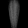 33999MC  7ymnC M: map data used to model this data C: citing same article ( |
|---|---|
| Similar structure data |

-Links
 PDBj
PDBj


- Assembly
| Deposited unit | 
|
|---|---|
| 1 |
|
-Components
| #1: Protein | Mass: 10318.810 Da / Num. of mol.: 6 Source method: isolated from a genetically manipulated source Source: (gene. exp.) Homo sapiens (human) / Gene: MAPT, MAPTL, MTBT1, TAU / Production host:  |
|---|
-Experimental details
-Experiment
| Experiment | Method: ELECTRON MICROSCOPY |
|---|---|
| EM experiment | Aggregation state: FILAMENT / 3D reconstruction method: helical reconstruction |
- Sample preparation
| Component | Name: Cryo-EM structure of amyloid fibril formed by tau (297-391) Type: ORGANELLE OR CELLULAR COMPONENT / Entity ID: all / Source: RECOMBINANT |
|---|---|
| Source (natural) | Organism: Homo sapiens (human) |
| Source (recombinant) | Organism: |
| Buffer solution | pH: 7.4 |
| Specimen | Embedding applied: NO / Shadowing applied: NO / Staining applied: NO / Vitrification applied: YES |
| Vitrification | Cryogen name: ETHANE |
- Electron microscopy imaging
| Experimental equipment |  Model: Titan Krios / Image courtesy: FEI Company |
|---|---|
| Microscopy | Model: FEI TITAN KRIOS |
| Electron gun | Electron source:  FIELD EMISSION GUN / Accelerating voltage: 300 kV / Illumination mode: FLOOD BEAM FIELD EMISSION GUN / Accelerating voltage: 300 kV / Illumination mode: FLOOD BEAM |
| Electron lens | Mode: BRIGHT FIELD / Nominal defocus max: 2200 nm / Nominal defocus min: 1400 nm |
| Image recording | Electron dose: 55 e/Å2 / Film or detector model: GATAN K3 (6k x 4k) |
- Processing
| CTF correction | Type: NONE |
|---|---|
| Helical symmerty | Angular rotation/subunit: 179.43 ° / Axial rise/subunit: 4.82 Å / Axial symmetry: C1 |
| 3D reconstruction | Resolution: 2.5 Å / Resolution method: FSC 0.143 CUT-OFF / Num. of particles: 354966 / Symmetry type: HELICAL |
