 Movie
Movie Controller

[English] 日本語
 Yorodumi
- PDB-7yp3: Crystal structure of elaiophylin glycosyltransferase in complex w... -
+ Open data

- Basic information
| Entry | Database: PDB / ID: 7yp3 | ||||||
|---|---|---|---|---|---|---|---|
| Title | Crystal structure of elaiophylin glycosyltransferase in complex with elaiophylin | ||||||
 Components Components | Glycosyltransferase | ||||||
 Keywords Keywords | TRANSFERASE / glycosyltransferase / elaiophylin / GT1 | ||||||
| Function / homology |  Function and homology information Function and homology informationUDP-glycosyltransferase activity / hexosyltransferase activity / antibiotic biosynthetic process / nucleotide binding Similarity search - Function | ||||||
| Biological species |  Streptomyces sp. SCSIO 01934 (bacteria) Streptomyces sp. SCSIO 01934 (bacteria) | ||||||
| Method |  X-RAY DIFFRACTION / SYNCHROTRON / MOLECULAR REPLACEMENT / Resolution: 2.1 Å X-RAY DIFFRACTION / SYNCHROTRON / MOLECULAR REPLACEMENT / Resolution: 2.1 Å | ||||||
 Authors Authors | Xu, T. / Liu, Q. / Gan, Q. / Liu, J. | ||||||
| Funding support |  China, 1items China, 1items
| ||||||
 Citation Citation | Journal: Acta Crystallogr D Struct Biol / Year: 2022 Title: Substrate-induced dimerization of elaiophylin glycosyltransferase reveals a novel self-activating form of glycosyltransferase for symmetric glycosylation. Authors: Xu, T. / Gan, Q. / Liu, Q. / Chen, R. / Zhen, X. / Zhang, C. / Liu, J. | ||||||
| History |
|
- Structure visualization
| Structure viewer | Molecule: MolmilJmol/JSmol |
|---|
- Downloads & links
-Download
| PDBx/mmCIF format | 7yp3.cif.gz | 588.4 KB | Display | PDBx/mmCIF format |
|---|---|---|---|---|
| PDB format | pdb7yp3.ent.gz | 486.6 KB | Display | PDB format |
| PDBx/mmJSON format | 7yp3.json.gz | Tree view | PDBx/mmJSON format | |
| Others |  Other downloads Other downloads |
-Validation report
| Arichive directory | https://data.pdbj.org/pub/pdb/validation_reports/yp/7yp3ftp://data.pdbj.org/pub/pdb/validation_reports/yp/7yp3 | HTTPS FTP |
|---|
-Related structure data
| Related structure data | 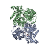 7yp4C 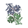 7yp5C 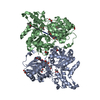 7yp6C 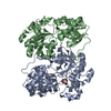 3d0rS S: Starting model for refinement C: citing same article ( |
|---|---|
| Similar structure data |
-Links
 PDBj
PDBj- Assembly
| Deposited unit | 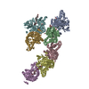
| ||||||||
|---|---|---|---|---|---|---|---|---|---|
| 1 | 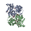
| ||||||||
| 2 | 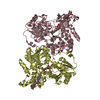
| ||||||||
| 3 | 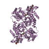
| ||||||||
| 4 | 
| ||||||||
| Unit cell |
|
-Components
| #1: Protein | Mass: 48544.961 Da / Num. of mol.: 7 Source method: isolated from a genetically manipulated source Source: (gene. exp.) Streptomyces sp. SCSIO 01934 (bacteria)Plasmid: pET28a / Production host: #2: Chemical | ChemComp-ACT /   Mass: 59.044 Da / Num. of mol.: 14 / Source method: obtained synthetically / Formula: C2H3O2 Mass: 59.044 Da / Num. of mol.: 14 / Source method: obtained synthetically / Formula: C2H3O2#3: Chemical | ChemComp-GOL /   Mass: 92.094 Da / Num. of mol.: 11 / Source method: obtained synthetically / Formula: C3H8O3 Mass: 92.094 Da / Num. of mol.: 11 / Source method: obtained synthetically / Formula: C3H8O3#4: Chemical | ChemComp-ELO / 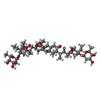  Mass: 1025.266 Da / Num. of mol.: 5 / Source method: obtained synthetically / Formula: C54H88O18 / Feature type: SUBJECT OF INVESTIGATION Mass: 1025.266 Da / Num. of mol.: 5 / Source method: obtained synthetically / Formula: C54H88O18 / Feature type: SUBJECT OF INVESTIGATION#5: Water | ChemComp-HOH / |  Mass: 18.015 Da / Num. of mol.: 959 / Source method: isolated from a natural source / Formula: H2O Mass: 18.015 Da / Num. of mol.: 959 / Source method: isolated from a natural source / Formula: H2OHas ligand of interest | Y | |
|---|
-Experimental details
-Experiment
| Experiment | Method: X-RAY DIFFRACTION / Number of used crystals: 1 |
|---|
- Sample preparation
| Crystal | Density Matthews: 2.84 Å3/Da / Density % sol: 56.64 % / Mosaicity: 0.54 ° |
|---|---|
| Crystal grow | Temperature: 293 K / Method: vapor diffusion / pH: 7.8 Details: 5% glycerol 3% PEG 3350 1.7M NH4OAC 0.1M Tris pH 7.8 5% Pentaerythritol ethoxylate |
-Data collection
| Diffraction | Mean temperature: 100 K / Serial crystal experiment: N | ||||||||||||||||||||||||||||||
|---|---|---|---|---|---|---|---|---|---|---|---|---|---|---|---|---|---|---|---|---|---|---|---|---|---|---|---|---|---|---|---|
| Diffraction source | Source: SYNCHROTRON / Site: SSRF / Beamline: BL17U / Wavelength: 0.97948 Å | ||||||||||||||||||||||||||||||
| Detector | Type: ADSC QUANTUM 315 / Detector: CCD / Date: Sep 29, 2010 | ||||||||||||||||||||||||||||||
| Radiation | Protocol: SINGLE WAVELENGTH / Monochromatic (M) / Laue (L): M / Scattering type: x-ray | ||||||||||||||||||||||||||||||
| Radiation wavelength | Wavelength: 0.97948 Å / Relative weight: 1 | ||||||||||||||||||||||||||||||
| Reflection | Resolution: 2.1→76.69 Å / Num. obs: 217985 / % possible obs: 98.7 % / Redundancy: 3 % / CC1/2: 0.996 / Rmerge(I) obs: 0.071 / Rpim(I) all: 0.048 / Rrim(I) all: 0.087 / Net I/σ(I): 8.3 / Num. measured all: 659590 / Scaling rejects: 1157 | ||||||||||||||||||||||||||||||
| Reflection shell | Diffraction-ID: 1
|
- Processing
| Software |
| |||||||||||||||||||||||||||||||||||||||||||||||||||||||
|---|---|---|---|---|---|---|---|---|---|---|---|---|---|---|---|---|---|---|---|---|---|---|---|---|---|---|---|---|---|---|---|---|---|---|---|---|---|---|---|---|---|---|---|---|---|---|---|---|---|---|---|---|---|---|---|---|
| Refinement | Method to determine structure: MOLECULAR REPLACEMENT Starting model: 3D0R Resolution: 2.1→76.01 Å / Cor.coef. Fo:Fc: 0.965 / Cor.coef. Fo:Fc free: 0.952 / SU B: 6.286 / SU ML: 0.149 / Cross valid method: THROUGHOUT / σ(F): 0 / ESU R: 0.188 / ESU R Free: 0.159 / Stereochemistry target values: MAXIMUM LIKELIHOOD Details: HYDROGENS HAVE BEEN ADDED IN THE RIDING POSITIONS U VALUES : REFINED INDIVIDUALLY
| |||||||||||||||||||||||||||||||||||||||||||||||||||||||
| Solvent computation | Ion probe radii: 0.8 Å / Shrinkage radii: 0.8 Å / VDW probe radii: 1.2 Å / Solvent model: MASK | |||||||||||||||||||||||||||||||||||||||||||||||||||||||
| Displacement parameters | Biso max: 198.49 Å2 / Biso mean: 53.986 Å2 / Biso min: 23.64 Å2
| |||||||||||||||||||||||||||||||||||||||||||||||||||||||
| Refinement step | Cycle: final / Resolution: 2.1→76.01 Å
| |||||||||||||||||||||||||||||||||||||||||||||||||||||||
| Refine LS restraints |
| |||||||||||||||||||||||||||||||||||||||||||||||||||||||
| LS refinement shell | Resolution: 2.1→2.155 Å / Rfactor Rfree error: 0 / Total num. of bins used: 20
|
