 Movie
Movie Controller

[English] 日本語
 Yorodumi
- PDB-7ym1: Structure of SsbA protein in complex with the anticancer drug 5-f... -
+ Open data

- Basic information
| Entry | Database: PDB / ID: 7ym1 | ||||||
|---|---|---|---|---|---|---|---|
| Title | Structure of SsbA protein in complex with the anticancer drug 5-fluorouracil | ||||||
 Components Components | Single-stranded DNA-binding protein | ||||||
 Keywords Keywords | DNA BINDING PROTEIN / single-strand DNA binding protein | ||||||
| Function / homology |  Function and homology information Function and homology informationnucleoid / single-stranded DNA binding / DNA recombination / DNA replication / DNA repair Similarity search - Function | ||||||
| Biological species |  Staphylococcus aureus subsp. aureus ED98 (bacteria) Staphylococcus aureus subsp. aureus ED98 (bacteria) | ||||||
| Method |  X-RAY DIFFRACTION / SYNCHROTRON / MOLECULAR REPLACEMENT / Resolution: 2.36 Å X-RAY DIFFRACTION / SYNCHROTRON / MOLECULAR REPLACEMENT / Resolution: 2.36 Å | ||||||
 Authors Authors | Huang, Y.H. / Yang, P.C. / Chiang, W.Y. / Lin, E.S. / Huang, C.Y. | ||||||
| Funding support | 1items
| ||||||
 Citation Citation | Journal: Int J Mol Sci / Year: 2023 Title: Crystal Structure of DNA Replication Protein SsbA Complexed with the Anticancer Drug 5-Fluorouracil. Authors: Su, H.H. / Huang, Y.H. / Lien, Y. / Yang, P.C. / Huang, C.Y. | ||||||
| History |
|
- Structure visualization
| Structure viewer | Molecule: MolmilJmol/JSmol |
|---|
- Downloads & links
-Download
| PDBx/mmCIF format | 7ym1.cif.gz | 54.7 KB | Display | PDBx/mmCIF format |
|---|---|---|---|---|
| PDB format | pdb7ym1.ent.gz | Display | PDB format | |
| PDBx/mmJSON format | 7ym1.json.gz | Tree view | PDBx/mmJSON format | |
| Others |  Other downloads Other downloads |
-Validation report
| Arichive directory | https://data.pdbj.org/pub/pdb/validation_reports/ym/7ym1ftp://data.pdbj.org/pub/pdb/validation_reports/ym/7ym1 | HTTPS FTP |
|---|
-Related structure data
| Related structure data | 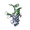 8gw5C  5xgtS S: Starting model for refinement C: citing same article ( |
|---|---|
| Similar structure data |
-Links
 PDBj
PDBj- Assembly
| Deposited unit | 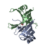
| ||||||||
|---|---|---|---|---|---|---|---|---|---|
| 1 |
| ||||||||
| Unit cell |
|
-Components
| #1: Protein | Mass: 12645.094 Da / Num. of mol.: 2 Source method: isolated from a genetically manipulated source Source: (gene. exp.) Staphylococcus aureus subsp. aureus ED98 (bacteria)Gene: ssb, ssb2, ssb_1, ssbA, A6762_01715, BN1321_80086, C7P97_01495, CSC87_00615, CV021_06850, DD547_00353, E3A28_06905, E3K14_01855, E4U00_06870, EDCC5055_00340, EP54_08250, EQ90_08535, G6Y24_ ...Gene: ssb, ssb2, ssb_1, ssbA, A6762_01715, BN1321_80086, C7P97_01495, CSC87_00615, CV021_06850, DD547_00353, E3A28_06905, E3K14_01855, E4U00_06870, EDCC5055_00340, EP54_08250, EQ90_08535, G6Y24_06705, GO782_08865, GO788_13850, GO793_13375, GO814_10580, GO941_16005, GO942_09280, GZ128_07070, GZ156_06265, HK402_02025, HMPREF3211_02451, HUW54_01845, NCTC10702_00704, NCTC5664_00336, NCTC7878_00400, NCTC7972_01579, QU38_04380, SA0759_00307, SA950122_00302, SAJPND4_00365, SAMEA70245418_01284 Production host: #2: Chemical | ChemComp-URF / | 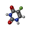  Mass: 130.077 Da / Num. of mol.: 1 / Source method: obtained synthetically / Formula: C4H3FN2O2 / Comment: medication, chemotherapy*YM Mass: 130.077 Da / Num. of mol.: 1 / Source method: obtained synthetically / Formula: C4H3FN2O2 / Comment: medication, chemotherapy*YM#3: Chemical | ChemComp-GOL / |   Mass: 92.094 Da / Num. of mol.: 1 / Source method: obtained synthetically / Formula: C3H8O3 / Feature type: SUBJECT OF INVESTIGATION Mass: 92.094 Da / Num. of mol.: 1 / Source method: obtained synthetically / Formula: C3H8O3 / Feature type: SUBJECT OF INVESTIGATION#4: Water | ChemComp-HOH / |  Mass: 18.015 Da / Num. of mol.: 52 / Source method: isolated from a natural source / Formula: H2O Mass: 18.015 Da / Num. of mol.: 52 / Source method: isolated from a natural source / Formula: H2OHas ligand of interest | Y | Has protein modification | N | |
|---|
-Experimental details
-Experiment
| Experiment | Method: X-RAY DIFFRACTION / Number of used crystals: 1 |
|---|
- Sample preparation
| Crystal | Density Matthews: 2.43 Å3/Da / Density % sol: 44.5 % |
|---|---|
| Crystal grow | Temperature: 298 K / Method: vapor diffusion, hanging drop / Details: 16% PEG4000, 100mM Tris, pH8.5, 200mM MgCl2 |
-Data collection
| Diffraction | Mean temperature: 298 K / Serial crystal experiment: N |
|---|---|
| Diffraction source | Source: SYNCHROTRON / Site: NSRRC  / Beamline: BL15A1 / Wavelength: 1 Å / Beamline: BL15A1 / Wavelength: 1 Å |
| Detector | Type: RAYONIX MX300-HS / Detector: CCD / Date: Apr 21, 2022 |
| Radiation | Protocol: SINGLE WAVELENGTH / Monochromatic (M) / Laue (L): M / Scattering type: x-ray |
| Radiation wavelength | Wavelength: 1 Å / Relative weight: 1 |
| Reflection | Resolution: 2.36→30 Å / Num. obs: 9854 / % possible obs: 100 % / Redundancy: 7.8 % / Rmerge(I) obs: 0.067 / Net I/σ(I): 31.6 |
| Reflection shell | Resolution: 2.36→2.44 Å / Redundancy: 7 % / Mean I/σ(I) obs: 8.2 / Num. unique obs: 964 / CC1/2: 0.974 / % possible all: 100 |
- Processing
| Software |
| ||||||||||||||||||||||||
|---|---|---|---|---|---|---|---|---|---|---|---|---|---|---|---|---|---|---|---|---|---|---|---|---|---|
| Refinement | Method to determine structure: MOLECULAR REPLACEMENT Starting model: 5XGT Resolution: 2.36→27.86 Å / SU ML: 0.25 / Cross valid method: THROUGHOUT / σ(F): 1.37 / Phase error: 24.57 / Stereochemistry target values: ML
| ||||||||||||||||||||||||
| Solvent computation | Shrinkage radii: 0.9 Å / VDW probe radii: 1.11 Å / Solvent model: FLAT BULK SOLVENT MODEL | ||||||||||||||||||||||||
| Displacement parameters | Biso max: 87.37 Å2 / Biso mean: 33.4772 Å2 / Biso min: 14.17 Å2 | ||||||||||||||||||||||||
| Refinement step | Cycle: final / Resolution: 2.36→27.86 Å
| ||||||||||||||||||||||||
| LS refinement shell | Refine-ID: X-RAY DIFFRACTION / Rfactor Rfree error: 0 / Total num. of bins used: 3 / % reflection obs: 100 %
|
