 Movie
Movie Controller

[English] 日本語
 Yorodumi
- PDB-7ylb: Two monobodies recognizing the conserved epitopes of SARS-CoV-2 N... -
+ Open data

- Basic information
| Entry | Database: PDB / ID: 7ylb | ||||||
|---|---|---|---|---|---|---|---|
| Title | Two monobodies recognizing the conserved epitopes of SARS-CoV-2 N antigen applicable to the broad COVID-19 diagnosis | ||||||
 Components Components |
| ||||||
 Keywords Keywords | VIRAL PROTEIN/IMMUNE SYSTEM / Engineered 10Fn3 / SARS-CoV-2 N / Monobody / VIRAL PROTEIN / VIRAL PROTEIN-IMMUNE SYSTEM complex | ||||||
| Function / homology |  Function and homology information Function and homology information: / response to host immune response / viral RNA genome packaging / negative regulation of interferon-beta production / poly(U) RNA binding / Maturation of nucleoprotein / intracellular membraneless organelle / positive regulation of NLRP3 inflammasome complex assembly / MHC class I protein binding / CD28 dependent PI3K/Akt signaling ...: / response to host immune response / viral RNA genome packaging / negative regulation of interferon-beta production / poly(U) RNA binding / Maturation of nucleoprotein / intracellular membraneless organelle / positive regulation of NLRP3 inflammasome complex assembly / MHC class I protein binding / CD28 dependent PI3K/Akt signaling / SARS-CoV-2 targets host intracellular signalling and regulatory pathways / protein sequestering activity / VEGFR2 mediated vascular permeability / molecular condensate scaffold activity / NOD1/2 Signaling Pathway / TAK1-dependent IKK and NF-kappa-B activation / MHC class I protein complex / DDX58/IFIH1-mediated induction of interferon-alpha/beta / RNA stem-loop binding / Interleukin-1 signaling / Interferon alpha/beta signaling / viral capsid / PIP3 activates AKT signaling / viral nucleocapsid / Transcription of SARS-CoV-2 sgRNAs / host cell endoplasmic reticulum-Golgi intermediate compartment / Translation of Structural Proteins / Virion Assembly and Release / host extracellular space / Induction of Cell-Cell Fusion / host cell Golgi apparatus / Attachment and Entry / host cell perinuclear region of cytoplasm / ribonucleoprotein complex / SARS-CoV-2 activates/modulates innate and adaptive immune responses / protein homodimerization activity / RNA binding / extracellular region / identical protein binding / cytoplasm Similarity search - Function | ||||||
| Biological species |   Severe acute respiratory syndrome coronavirus 2 Severe acute respiratory syndrome coronavirus 2 Homo sapiens (human) Homo sapiens (human) | ||||||
| Method |  X-RAY DIFFRACTION / SYNCHROTRON / MOLECULAR REPLACEMENT / Resolution: 2.41 Å X-RAY DIFFRACTION / SYNCHROTRON / MOLECULAR REPLACEMENT / Resolution: 2.41 Å | ||||||
 Authors Authors | Hu, M. / Du, Y. / Sun, R. / Hao, Q. | ||||||
| Funding support | 1items
| ||||||
 Citation Citation | Journal: To Be Published Title: Two monobodies recognizing the conserved epitopes of SARS-CoV-2 N antigen applicable to the broad COVID-19 diagnosis Authors: Hu, M. / Du, Y. / Sun, R. / Hao, Q. | ||||||
| History |
|
- Structure visualization
| Structure viewer | Molecule: MolmilJmol/JSmol |
|---|
- Downloads & links
-Download
| PDBx/mmCIF format | 7ylb.cif.gz | 246.8 KB | Display | PDBx/mmCIF format |
|---|---|---|---|---|
| PDB format | pdb7ylb.ent.gz | 198.8 KB | Display | PDB format |
| PDBx/mmJSON format | 7ylb.json.gz | Tree view | PDBx/mmJSON format | |
| Others |  Other downloads Other downloads |
-Validation report
| Arichive directory | https://data.pdbj.org/pub/pdb/validation_reports/yl/7ylbftp://data.pdbj.org/pub/pdb/validation_reports/yl/7ylb | HTTPS FTP |
|---|
-Related structure data
| Related structure data |  7yldC  1fnaS 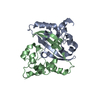 6yunS S: Starting model for refinement C: citing same article ( |
|---|---|
| Similar structure data |
-Links
 PDBj
PDBj


- Assembly
| Deposited unit | 
| ||||||||
|---|---|---|---|---|---|---|---|---|---|
| 1 | 
| ||||||||
| 2 | 
| ||||||||
| 3 | 
| ||||||||
| 4 | 
| ||||||||
| Unit cell |
|
-Components
| #1: Protein | Mass: 13562.218 Da / Num. of mol.: 8 / Fragment: CTD Source method: isolated from a genetically manipulated source Source: (gene. exp.) Severe acute respiratory syndrome coronavirus 2Production host:  #2: Protein | Mass: 10384.603 Da / Num. of mol.: 4 Source method: isolated from a genetically manipulated source Source: (gene. exp.) Homo sapiens (human) / Production host: #3: Chemical | ChemComp-SO4 /   Mass: 96.063 Da / Num. of mol.: 4 / Source method: obtained synthetically / Formula: SO4 Mass: 96.063 Da / Num. of mol.: 4 / Source method: obtained synthetically / Formula: SO4Has ligand of interest | N | |
|---|
-Experimental details
-Experiment
| Experiment | Method: X-RAY DIFFRACTION / Number of used crystals: 1 |
|---|
- Sample preparation
| Crystal | Density Matthews: 3.19 Å3/Da / Density % sol: 61.5 % |
|---|---|
| Crystal grow | Temperature: 293.15 K / Method: evaporation / Details: 0.2M K2SO4, 18% PEG 3350 |
-Data collection
| Diffraction | Mean temperature: 100 K / Serial crystal experiment: N |
|---|---|
| Diffraction source | Source: SYNCHROTRON / Site: SSRF  / Beamline: BL19U1 / Wavelength: 0.979 Å / Beamline: BL19U1 / Wavelength: 0.979 Å |
| Detector | Type: DECTRIS PILATUS3 6M / Detector: PIXEL / Date: Dec 5, 2020 |
| Radiation | Protocol: SINGLE WAVELENGTH / Monochromatic (M) / Laue (L): M / Scattering type: x-ray |
| Radiation wavelength | Wavelength: 0.979 Å / Relative weight: 1 |
| Reflection | Resolution: 2.41→138.261 Å / Num. obs: 74632 / % possible obs: 99.8 % / Redundancy: 6.6 % / Rmerge(I) obs: 0.186 / Net I/σ(I): 7.6 |
| Reflection shell | Resolution: 2.41→2.45 Å / Rmerge(I) obs: 0.945 / Num. unique obs: 3673 |
- Processing
| Software |
| |||||||||||||||||||||||||||||||||||||||||||||
|---|---|---|---|---|---|---|---|---|---|---|---|---|---|---|---|---|---|---|---|---|---|---|---|---|---|---|---|---|---|---|---|---|---|---|---|---|---|---|---|---|---|---|---|---|---|---|
| Refinement | Method to determine structure: MOLECULAR REPLACEMENT Starting model: 6YUN, 1FNA Resolution: 2.41→138.261 Å / Cor.coef. Fo:Fc: 0.889 / Cor.coef. Fo:Fc free: 0.821 / SU B: 12.799 / SU ML: 0.293 / Cross valid method: THROUGHOUT / σ(F): 0 / ESU R: 0.361 / ESU R Free: 0.296 / Stereochemistry target values: MAXIMUM LIKELIHOOD / Details: U VALUES : REFINED INDIVIDUALLY
| |||||||||||||||||||||||||||||||||||||||||||||
| Solvent computation | Ion probe radii: 0.8 Å / Shrinkage radii: 0.8 Å / VDW probe radii: 1.2 Å / Solvent model: MASK | |||||||||||||||||||||||||||||||||||||||||||||
| Displacement parameters | Biso max: 137.72 Å2 / Biso mean: 41.358 Å2 / Biso min: 15.18 Å2
| |||||||||||||||||||||||||||||||||||||||||||||
| Refinement step | Cycle: final / Resolution: 2.41→138.261 Å
| |||||||||||||||||||||||||||||||||||||||||||||
| Refine LS restraints |
| |||||||||||||||||||||||||||||||||||||||||||||
| LS refinement shell | Resolution: 2.41→2.468 Å / Rfactor Rfree error: 0
|
