 Movie
Movie Controller

[English] 日本語
 Yorodumi
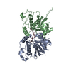
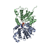
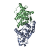
- PDB-7yhl: Crystal Structure of the ring nuclease Sso2081 from Saccharolobus... -
+ Open data

- Basic information
| Entry | Database: PDB / ID: 7yhl | ||||||
|---|---|---|---|---|---|---|---|
| Title | Crystal Structure of the ring nuclease Sso2081 from Saccharolobus solfataricus in complex with free phosphate | ||||||
 Components Components | CRISPR system ring nuclease SSO2081 | ||||||
 Keywords Keywords | HYDROLASE / Sso2081 / ring nuclease / wild type / free phosphate | ||||||
| Function / homology | CRISPR-assoc protein, NE0113/Csx13 / CRISPR-associated protein NE0113 (Cas_NE0113) / Restriction endonuclease type II-like / Lyases; Phosphorus-oxygen lyases / defense response to virus / lyase activity / cytoplasm / PHOSPHATE ION / CRISPR system ring nuclease SSO2081 Function and homology information Function and homology information | ||||||
| Biological species |   Saccharolobus solfataricus P2 (archaea) Saccharolobus solfataricus P2 (archaea) | ||||||
| Method |  X-RAY DIFFRACTION / SYNCHROTRON / SAD / Resolution: 2.7 Å X-RAY DIFFRACTION / SYNCHROTRON / SAD / Resolution: 2.7 Å | ||||||
 Authors Authors | Lin, Z. / Du, L. / Luo, Z. | ||||||
| Funding support |  China, 1items China, 1items
| ||||||
 Citation Citation | Journal: Nucleic Acids Res. / Year: 2023 Title: Molecular basis of stepwise cyclic tetra-adenylate cleavage by the type III CRISPR ring nuclease Crn1/Sso2081. Authors: Du, L. / Zhang, D. / Luo, Z. / Lin, Z. | ||||||
| History |
|
- Structure visualization
| Structure viewer | Molecule: MolmilJmol/JSmol |
|---|
- Downloads & links
-Download
| PDBx/mmCIF format | 7yhl.cif.gz | 193.4 KB | Display | PDBx/mmCIF format |
|---|---|---|---|---|
| PDB format | pdb7yhl.ent.gz | 121 KB | Display | PDB format |
| PDBx/mmJSON format | 7yhl.json.gz | Tree view | PDBx/mmJSON format | |
| Others |  Other downloads Other downloads |
-Validation report
| Summary document | 7yhl_validation.pdf.gz | 457.1 KB | Display | wwPDB validaton report |
|---|---|---|---|---|
| Full document | 7yhl_full_validation.pdf.gz | 463.9 KB | Display | |
| Data in XML | 7yhl_validation.xml.gz | 14.6 KB | Display | |
| Data in CIF | 7yhl_validation.cif.gz | 18.7 KB | Display | |
| Arichive directory | https://data.pdbj.org/pub/pdb/validation_reports/yh/7yhlftp://data.pdbj.org/pub/pdb/validation_reports/yh/7yhl | HTTPS FTP |
-Related structure data
-Links
 PDBj
PDBj


- Assembly
| Deposited unit | 
| ||||||||
|---|---|---|---|---|---|---|---|---|---|
| 1 |
| ||||||||
| Unit cell |
|
-Components
| #1: Protein | Mass: 20431.061 Da / Num. of mol.: 2 Source method: isolated from a genetically manipulated source Source: (gene. exp.) Saccharolobus solfataricus P2 (archaea)Strain: ATCC 35092 / DSM 1617 / JCM 11322 / P2 / Gene: SSO2081 / Production host:  References: UniProt: Q7LYJ6, Lyases; Phosphorus-oxygen lyases #2: Chemical |   Mass: 94.971 Da / Num. of mol.: 2 / Source method: obtained synthetically / Formula: PO4 Mass: 94.971 Da / Num. of mol.: 2 / Source method: obtained synthetically / Formula: PO4#3: Water | ChemComp-HOH / |  Mass: 18.015 Da / Num. of mol.: 3 / Source method: isolated from a natural source / Formula: H2O Mass: 18.015 Da / Num. of mol.: 3 / Source method: isolated from a natural source / Formula: H2OHas ligand of interest | N | Has protein modification | Y | |
|---|
-Experimental details
-Experiment
| Experiment | Method: X-RAY DIFFRACTION / Number of used crystals: 1 |
|---|
- Sample preparation
| Crystal | Density Matthews: 2.13 Å3/Da / Density % sol: 42.17 % |
|---|---|
| Crystal grow | Temperature: 298 K / Method: vapor diffusion Details: 0.1 M Sodium acetate trihydrate, pH 4.5, 25% w/v Polyethylene glycol 1500 |
-Data collection
| Diffraction | Mean temperature: 100 K / Serial crystal experiment: N | ||||||||||||||||||
|---|---|---|---|---|---|---|---|---|---|---|---|---|---|---|---|---|---|---|---|
| Diffraction source | Source: SYNCHROTRON / Site: SSRF / Beamline: BL02U1 / Wavelength: 0.97919 Å | ||||||||||||||||||
| Detector | Type: DECTRIS EIGER2 S 9M / Detector: PIXEL / Date: Jan 1, 2022 | ||||||||||||||||||
| Radiation | Protocol: SINGLE WAVELENGTH / Monochromatic (M) / Laue (L): M / Scattering type: x-ray | ||||||||||||||||||
| Radiation wavelength | Wavelength: 0.97919 Å / Relative weight: 1 | ||||||||||||||||||
| Reflection twin |
| ||||||||||||||||||
| Reflection | Resolution: 2.7→40.95 Å / Num. obs: 9099 / % possible obs: 95.55 % / Redundancy: 5.29 % / CC1/2: 0.998 / Rmerge(I) obs: 0.067 / Net I/σ(I): 10.9 | ||||||||||||||||||
| Reflection shell | Resolution: 2.7→2.79 Å / Redundancy: 5.32 % / Rmerge(I) obs: 0.748 / Mean I/σ(I) obs: 1.5 / Num. unique obs: 924 / CC1/2: 0.805 / % possible all: 95.55 |
- Processing
| Software |
| |||||||||||||||||||||||||||||||||||||||||||||||||||||||||||||||||||||||||||||||||||||||||||||||||||||||||||||||||||||||||||||||||||||||||||||||||||||||||||
|---|---|---|---|---|---|---|---|---|---|---|---|---|---|---|---|---|---|---|---|---|---|---|---|---|---|---|---|---|---|---|---|---|---|---|---|---|---|---|---|---|---|---|---|---|---|---|---|---|---|---|---|---|---|---|---|---|---|---|---|---|---|---|---|---|---|---|---|---|---|---|---|---|---|---|---|---|---|---|---|---|---|---|---|---|---|---|---|---|---|---|---|---|---|---|---|---|---|---|---|---|---|---|---|---|---|---|---|---|---|---|---|---|---|---|---|---|---|---|---|---|---|---|---|---|---|---|---|---|---|---|---|---|---|---|---|---|---|---|---|---|---|---|---|---|---|---|---|---|---|---|---|---|---|---|---|---|
| Refinement | Method to determine structure: SAD / Resolution: 2.7→40.95 Å / Cor.coef. Fo:Fc: 0.942 / Cor.coef. Fo:Fc free: 0.927 / SU B: 47.161 / SU ML: 0.451 / Cross valid method: THROUGHOUT / ESU R Free: 0.453 Details: Hydrogens have been added in their riding positions
| |||||||||||||||||||||||||||||||||||||||||||||||||||||||||||||||||||||||||||||||||||||||||||||||||||||||||||||||||||||||||||||||||||||||||||||||||||||||||||
| Solvent computation | Ion probe radii: 0.8 Å / Shrinkage radii: 0.8 Å / VDW probe radii: 1.2 Å / Solvent model: MASK BULK SOLVENT | |||||||||||||||||||||||||||||||||||||||||||||||||||||||||||||||||||||||||||||||||||||||||||||||||||||||||||||||||||||||||||||||||||||||||||||||||||||||||||
| Displacement parameters | Biso mean: 117.165 Å2
| |||||||||||||||||||||||||||||||||||||||||||||||||||||||||||||||||||||||||||||||||||||||||||||||||||||||||||||||||||||||||||||||||||||||||||||||||||||||||||
| Refinement step | Cycle: LAST / Resolution: 2.7→40.95 Å
| |||||||||||||||||||||||||||||||||||||||||||||||||||||||||||||||||||||||||||||||||||||||||||||||||||||||||||||||||||||||||||||||||||||||||||||||||||||||||||
| Refine LS restraints |
| |||||||||||||||||||||||||||||||||||||||||||||||||||||||||||||||||||||||||||||||||||||||||||||||||||||||||||||||||||||||||||||||||||||||||||||||||||||||||||
| LS refinement shell |
| |||||||||||||||||||||||||||||||||||||||||||||||||||||||||||||||||||||||||||||||||||||||||||||||||||||||||||||||||||||||||||||||||||||||||||||||||||||||||||
| Refinement TLS params. | Method: refined / Refine-ID: X-RAY DIFFRACTION
| |||||||||||||||||||||||||||||||||||||||||||||||||||||||||||||||||||||||||||||||||||||||||||||||||||||||||||||||||||||||||||||||||||||||||||||||||||||||||||
| Refinement TLS group | Selection: ALL |
