 Movie
Movie Controller

[English] 日本語
 Yorodumi
- PDB-7yh4: Crystal structure of human cytosolic beta-alanyl lysine dipeptida... -
+ Open data

- Basic information
| Entry | Database: PDB / ID: 7yh4 | ||||||
|---|---|---|---|---|---|---|---|
| Title | Crystal structure of human cytosolic beta-alanyl lysine dipeptidase (PM20D2) | ||||||
 Components Components | Xaa-Arg dipeptidase | ||||||
 Keywords Keywords | HYDROLASE / Dipeptidase / carnosine proofreading / DNA repair | ||||||
| Function / homology |  Function and homology information Function and homology informationXaa-Arg dipeptidase / regulation of protein metabolic process / dipeptidase activity / carboxypeptidase activity / proteolysis / nucleoplasm / identical protein binding Similarity search - Function | ||||||
| Biological species |  Homo sapiens (human) Homo sapiens (human) | ||||||
| Method |  X-RAY DIFFRACTION / SYNCHROTRON / MOLECULAR REPLACEMENT / Resolution: 2.03 Å X-RAY DIFFRACTION / SYNCHROTRON / MOLECULAR REPLACEMENT / Resolution: 2.03 Å | ||||||
 Authors Authors | Chandravanshi, K. / Gaur, N.K. / Kumar, A. / Makde, R.D. | ||||||
| Funding support |  India, 1items India, 1items
| ||||||
 Citation Citation | Journal: To Be Published Title: Crystal structure of human cytosolic beta-alanyl lysine dipeptidase (PM20D2) Authors: Chandravanshi, K. / Gaur, N.K. / Kumar, A. / Makde, R.D. | ||||||
| History |
|
- Structure visualization
| Structure viewer | Molecule: MolmilJmol/JSmol |
|---|
- Downloads & links
-Download
| PDBx/mmCIF format | 7yh4.cif.gz | 174.9 KB | Display | PDBx/mmCIF format |
|---|---|---|---|---|
| PDB format | pdb7yh4.ent.gz | 135.8 KB | Display | PDB format |
| PDBx/mmJSON format | 7yh4.json.gz | Tree view | PDBx/mmJSON format | |
| Others |  Other downloads Other downloads |
-Validation report
| Arichive directory | https://data.pdbj.org/pub/pdb/validation_reports/yh/7yh4ftp://data.pdbj.org/pub/pdb/validation_reports/yh/7yh4 | HTTPS FTP |
|---|
-Related structure data
| Related structure data | 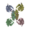 3ramS S: Starting model for refinement |
|---|---|
| Similar structure data |
-Links
 PDBj
PDBj
- Assembly
| Deposited unit | 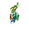
| ||||||||
|---|---|---|---|---|---|---|---|---|---|
| 1 | 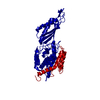
| ||||||||
| Unit cell |
| ||||||||
| Components on special symmetry positions |
|
-Components
| #1: Protein | Mass: 49748.809 Da / Num. of mol.: 1 Source method: isolated from a genetically manipulated source Source: (gene. exp.) Homo sapiens (human) / Gene: PM20D2, ACY1L2 / Plasmid: PST50STRN / Details (production host): T7-based / Production host:  | ||||||
|---|---|---|---|---|---|---|---|
| #2: Chemical |   Mass: 65.409 Da / Num. of mol.: 2 / Source method: obtained synthetically / Formula: Zn / Feature type: SUBJECT OF INVESTIGATION Mass: 65.409 Da / Num. of mol.: 2 / Source method: obtained synthetically / Formula: Zn / Feature type: SUBJECT OF INVESTIGATION#3: Water | ChemComp-HOH / |  Mass: 18.015 Da / Num. of mol.: 95 / Source method: isolated from a natural source / Formula: H2O Mass: 18.015 Da / Num. of mol.: 95 / Source method: isolated from a natural source / Formula: H2OHas ligand of interest | Y | Has protein modification | N | |
-Experimental details
-Experiment
| Experiment | Method: X-RAY DIFFRACTION / Number of used crystals: 1 |
|---|
- Sample preparation
| Crystal | Density Matthews: 2.76 Å3/Da / Density % sol: 56.06 % / Description: Hexagonal |
|---|---|
| Crystal grow | Temperature: 294 K / Method: microbatch / pH: 4.6 Details: 70 mM calcium chloride, 35 mM sodium acetate pH4.6, 7% isopropanol, 15% glycerol, 20mM Tris-Cl, 200 mM NaCl (mixed in 1:1) |
-Data collection
| Diffraction | Mean temperature: 100 K / Serial crystal experiment: Y |
|---|---|
| Diffraction source | Source: SYNCHROTRON / Site: RRCAT INDUS-2 / Beamline: PX-BL21 / Wavelength: 0.987 Å |
| Detector | Type: MARMOSAIC 225 mm CCD / Detector: CCD / Date: Aug 13, 2021 / Details: mirror |
| Radiation | Protocol: SINGLE WAVELENGTH / Monochromatic (M) / Laue (L): M / Scattering type: x-ray |
| Radiation wavelength | Wavelength: 0.987 Å / Relative weight: 1 |
| Reflection | Resolution: 2.03→44.59 Å / Num. obs: 35868 / % possible obs: 100 % / Redundancy: 7.2 % / Biso Wilson estimate: 42.23 Å2 / CC1/2: 1 / Rmerge(I) obs: 0.049 / Rpim(I) all: 0.02 / Rrim(I) all: 0.053 / Net I/σ(I): 23.3 |
| Reflection shell | Resolution: 2.03→2.08 Å / Redundancy: 5.4 % / Rmerge(I) obs: 1.14 / Mean I/σ(I) obs: 1.7 / Num. unique obs: 2616 / CC1/2: 0.69 / Rpim(I) all: 0.54 / % possible all: 99.8 |
| Serial crystallography sample delivery | Method: fixed target |
- Processing
| Software |
| ||||||||||||||||||||||||||||||||||||||||||||||||||||||||||||||||||||||||||||||||||||||||||||||||||
|---|---|---|---|---|---|---|---|---|---|---|---|---|---|---|---|---|---|---|---|---|---|---|---|---|---|---|---|---|---|---|---|---|---|---|---|---|---|---|---|---|---|---|---|---|---|---|---|---|---|---|---|---|---|---|---|---|---|---|---|---|---|---|---|---|---|---|---|---|---|---|---|---|---|---|---|---|---|---|---|---|---|---|---|---|---|---|---|---|---|---|---|---|---|---|---|---|---|---|---|
| Refinement | Method to determine structure: MOLECULAR REPLACEMENT Starting model: 3RAM Resolution: 2.03→35.624 Å / SU ML: 0.21 / Cross valid method: THROUGHOUT / σ(F): 1.33 / Phase error: 26.51 / Stereochemistry target values: ML
| ||||||||||||||||||||||||||||||||||||||||||||||||||||||||||||||||||||||||||||||||||||||||||||||||||
| Solvent computation | Shrinkage radii: 0.9 Å / VDW probe radii: 1.11 Å / Solvent model: FLAT BULK SOLVENT MODEL | ||||||||||||||||||||||||||||||||||||||||||||||||||||||||||||||||||||||||||||||||||||||||||||||||||
| Refinement step | Cycle: LAST / Resolution: 2.03→35.624 Å
| ||||||||||||||||||||||||||||||||||||||||||||||||||||||||||||||||||||||||||||||||||||||||||||||||||
| Refine LS restraints |
| ||||||||||||||||||||||||||||||||||||||||||||||||||||||||||||||||||||||||||||||||||||||||||||||||||
| LS refinement shell |
| ||||||||||||||||||||||||||||||||||||||||||||||||||||||||||||||||||||||||||||||||||||||||||||||||||
| Refinement TLS params. | Method: refined / Origin x: 7.628 Å / Origin y: 21.1855 Å / Origin z: 28.8788 Å
| ||||||||||||||||||||||||||||||||||||||||||||||||||||||||||||||||||||||||||||||||||||||||||||||||||
| Refinement TLS group | Selection details: all |
