 Movie
Movie Controller


+ Open data

- Basic information
| Entry | Database: PDB / ID: 7ygw | |||||||||
|---|---|---|---|---|---|---|---|---|---|---|
| Title | Crystal structure of the Zn2+-bound EFhd1/Swiprosin-2 | |||||||||
 Components Components | EF-hand domain-containing protein D1 | |||||||||
 Keywords Keywords | METAL BINDING PROTEIN / EF-hand containing protein / mitochondria | |||||||||
| Function / homology |  Function and homology information Function and homology informationregulation of cellular hyperosmotic salinity response / calcium ion sensor activity / neuron projection development / mitochondrial inner membrane / calcium ion binding / mitochondrion Similarity search - Function | |||||||||
| Biological species |  | |||||||||
| Method |  X-RAY DIFFRACTION / SYNCHROTRON / MOLECULAR REPLACEMENT / Resolution: 1.72 Å X-RAY DIFFRACTION / SYNCHROTRON / MOLECULAR REPLACEMENT / Resolution: 1.72 Å | |||||||||
 Authors Authors | Mun, S.A. / Park, J. / Kang, J.Y. / Park, T. / Jin, M. / Yang, J. / Eom, S.H. | |||||||||
| Funding support |  Korea, Republic Of, 2items Korea, Republic Of, 2items
| |||||||||
 Citation Citation | Journal: Iucrj / Year: 2023 Title: Structural and biochemical insights into Zn 2+ -bound EF-hand proteins, EFhd1 and EFhd2. Authors: Mun, S.A. / Park, J. / Kang, J.Y. / Park, T. / Jin, M. / Yang, J. / Eom, S.H. | |||||||||
| History |
|
- Structure visualization
| Structure viewer | Molecule: MolmilJmol/JSmol |
|---|
- Downloads & links
-Download
| PDBx/mmCIF format | 7ygw.cif.gz | 42.7 KB | Display | PDBx/mmCIF format |
|---|---|---|---|---|
| PDB format | pdb7ygw.ent.gz | 22.6 KB | Display | PDB format |
| PDBx/mmJSON format | 7ygw.json.gz | Tree view | PDBx/mmJSON format | |
| Others |  Other downloads Other downloads |
-Validation report
| Summary document | 7ygw_validation.pdf.gz | 2.5 MB | Display | wwPDB validaton report |
|---|---|---|---|---|
| Full document | 7ygw_full_validation.pdf.gz | 2.5 MB | Display | |
| Data in XML | 7ygw_validation.xml.gz | 6.3 KB | Display | |
| Data in CIF | 7ygw_validation.cif.gz | 7.7 KB | Display | |
| Arichive directory | https://data.pdbj.org/pub/pdb/validation_reports/yg/7ygwftp://data.pdbj.org/pub/pdb/validation_reports/yg/7ygw | HTTPS FTP |
-Related structure data
| Related structure data | 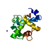 7ygvC  7ygyC 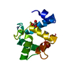 7cltS S: Starting model for refinement C: citing same article ( |
|---|---|
| Similar structure data |
-Links
 PDBj
PDBj- Assembly
| Deposited unit | 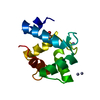
| ||||||||||||
|---|---|---|---|---|---|---|---|---|---|---|---|---|---|
| 1 |
| ||||||||||||
| Unit cell |
|
-Components
| #1: Protein | Mass: 14592.832 Da / Num. of mol.: 1 Source method: isolated from a genetically manipulated source Details: [GAMGS + EFhd1(69-193)](GAMGS: the cleaved site after GST-TEV tag cleavage) Source: (gene. exp.)  | ||||||
|---|---|---|---|---|---|---|---|
| #2: Chemical | ChemComp-ZN /   Mass: 65.409 Da / Num. of mol.: 4 / Source method: obtained synthetically / Formula: Zn / Feature type: SUBJECT OF INVESTIGATION Mass: 65.409 Da / Num. of mol.: 4 / Source method: obtained synthetically / Formula: Zn / Feature type: SUBJECT OF INVESTIGATION#3: Chemical | ChemComp-GOL / |   Mass: 92.094 Da / Num. of mol.: 1 / Source method: obtained synthetically / Formula: C3H8O3 / Feature type: SUBJECT OF INVESTIGATION Mass: 92.094 Da / Num. of mol.: 1 / Source method: obtained synthetically / Formula: C3H8O3 / Feature type: SUBJECT OF INVESTIGATION#4: Water | ChemComp-HOH / |  Mass: 18.015 Da / Num. of mol.: 32 / Source method: isolated from a natural source / Formula: H2O Mass: 18.015 Da / Num. of mol.: 32 / Source method: isolated from a natural source / Formula: H2OHas ligand of interest | Y | |
-Experimental details
-Experiment
| Experiment | Method: X-RAY DIFFRACTION / Number of used crystals: 1 |
|---|
- Sample preparation
| Crystal | Density Matthews: 2.26 Å3/Da / Density % sol: 45.6 % |
|---|---|
| Crystal grow | Temperature: 293 K / Method: vapor diffusion, sitting drop / pH: 8.5 Details: 0.1 M Tris-HCl pH 8.5, 2.5 mM ZnSO4, 25% (w/v) Jeffamine ED-2001 |
-Data collection
| Diffraction | Mean temperature: 100 K / Serial crystal experiment: N |
|---|---|
| Diffraction source | Source: SYNCHROTRON / Site: PAL/PLS / Beamline: 5C (4A) / Wavelength: 1.28255 Å |
| Detector | Type: DECTRIS EIGER X 9M / Detector: PIXEL / Date: Jun 3, 2020 |
| Radiation | Protocol: SINGLE WAVELENGTH / Monochromatic (M) / Laue (L): M / Scattering type: x-ray |
| Radiation wavelength | Wavelength: 1.28255 Å / Relative weight: 1 |
| Reflection | Resolution: 1.72→50 Å / Num. obs: 14577 / % possible obs: 99.5 % / Redundancy: 12.5 % / Biso Wilson estimate: 19.1 Å2 / CC1/2: 1 / Rmerge(I) obs: 0.079 / Net I/σ(I): 24.4 |
| Reflection shell | Resolution: 1.72→1.75 Å / Num. unique obs: 685 / CC1/2: 0.774 / % possible all: 94.5 |
- Processing
| Software |
| ||||||||||||||||||||||||||||||||||||||||||||||||||||||||||||||||||||||||||||||||||||||||||||||||||||||||||||||||||||||||||||||||||||||||||||
|---|---|---|---|---|---|---|---|---|---|---|---|---|---|---|---|---|---|---|---|---|---|---|---|---|---|---|---|---|---|---|---|---|---|---|---|---|---|---|---|---|---|---|---|---|---|---|---|---|---|---|---|---|---|---|---|---|---|---|---|---|---|---|---|---|---|---|---|---|---|---|---|---|---|---|---|---|---|---|---|---|---|---|---|---|---|---|---|---|---|---|---|---|---|---|---|---|---|---|---|---|---|---|---|---|---|---|---|---|---|---|---|---|---|---|---|---|---|---|---|---|---|---|---|---|---|---|---|---|---|---|---|---|---|---|---|---|---|---|---|---|---|
| Refinement | Method to determine structure: MOLECULAR REPLACEMENT Starting model: 7CLT Resolution: 1.72→37.96 Å / SU ML: 0.1872 / Cross valid method: FREE R-VALUE / σ(F): 1.37 / Phase error: 20.9894 Stereochemistry target values: GeoStd + Monomer Library + CDL v1.2
| ||||||||||||||||||||||||||||||||||||||||||||||||||||||||||||||||||||||||||||||||||||||||||||||||||||||||||||||||||||||||||||||||||||||||||||
| Solvent computation | Shrinkage radii: 0.9 Å / VDW probe radii: 1.11 Å / Solvent model: FLAT BULK SOLVENT MODEL | ||||||||||||||||||||||||||||||||||||||||||||||||||||||||||||||||||||||||||||||||||||||||||||||||||||||||||||||||||||||||||||||||||||||||||||
| Displacement parameters | Biso mean: 22.36 Å2 | ||||||||||||||||||||||||||||||||||||||||||||||||||||||||||||||||||||||||||||||||||||||||||||||||||||||||||||||||||||||||||||||||||||||||||||
| Refinement step | Cycle: LAST / Resolution: 1.72→37.96 Å
| ||||||||||||||||||||||||||||||||||||||||||||||||||||||||||||||||||||||||||||||||||||||||||||||||||||||||||||||||||||||||||||||||||||||||||||
| Refine LS restraints |
| ||||||||||||||||||||||||||||||||||||||||||||||||||||||||||||||||||||||||||||||||||||||||||||||||||||||||||||||||||||||||||||||||||||||||||||
| LS refinement shell |
|
