[English] 日本語
Yorodumi
- PDB-7yco: Crystal structure of SARS-CoV-2 Receptor Binding Domain bound to ... -

+
Open data


ID or keywords:

Loading...

-
Basic information

Entry
Database: PDB / ID: 7yco
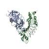
TitleCrystal structure of SARS-CoV-2 Receptor Binding Domain bound to A6 repebody
Components
  • Repebody (A6)
  • Spike protein S1
KeywordsPROTEIN BINDING / Severe acute respiratory syndrome coronavirus 2 / Complex
Function / homology
Function and homology information


symbiont-mediated disruption of host tissue / Maturation of spike protein / host cell surface / Translation of Structural Proteins / Virion Assembly and Release / Lectin pathway of complement activation / host extracellular region / symbiont-mediated-mediated suppression of host tetherin activity / structural constituent of virion / Induction of Cell-Cell Fusion ...symbiont-mediated disruption of host tissue / Maturation of spike protein / host cell surface / Translation of Structural Proteins / Virion Assembly and Release / Lectin pathway of complement activation / host extracellular region / symbiont-mediated-mediated suppression of host tetherin activity / structural constituent of virion / Induction of Cell-Cell Fusion / positive regulation of viral entry into host cell / Initial triggering of complement / membrane fusion / host cell endoplasmic reticulum-Golgi intermediate compartment membrane / Attachment and Entry / entry receptor-mediated virion attachment to host cell / receptor-mediated virion attachment to host cell / host cell surface receptor binding / symbiont-mediated suppression of host innate immune response / endocytosis involved in viral entry into host cell / receptor ligand activity / fusion of virus membrane with host plasma membrane / fusion of virus membrane with host endosome membrane / viral envelope / symbiont entry into host cell / virion attachment to host cell / host cell plasma membrane / SARS-CoV-2 activates/modulates innate and adaptive immune responses / virion membrane / membrane / identical protein binding / plasma membrane
Similarity search - Function
Spike (S) protein S1 subunit, receptor-binding domain, SARS-CoV-2 / Spike (S) protein S1 subunit, N-terminal domain, SARS-CoV-like / Coronavirus spike glycoprotein S1, C-terminal / Coronavirus spike glycoprotein S1, C-terminal / Spike glycoprotein, N-terminal domain superfamily / Spike S1 subunit, receptor binding domain superfamily, betacoronavirus / Spike glycoprotein, betacoronavirus / Betacoronavirus spike (S) glycoprotein S1 subunit N-terminal (NTD) domain profile. / Spike glycoprotein S1, N-terminal domain, betacoronavirus-like / Betacoronavirus-like spike glycoprotein S1, N-terminal ...Spike (S) protein S1 subunit, receptor-binding domain, SARS-CoV-2 / Spike (S) protein S1 subunit, N-terminal domain, SARS-CoV-like / Coronavirus spike glycoprotein S1, C-terminal / Coronavirus spike glycoprotein S1, C-terminal / Spike glycoprotein, N-terminal domain superfamily / Spike S1 subunit, receptor binding domain superfamily, betacoronavirus / Spike glycoprotein, betacoronavirus / Betacoronavirus spike (S) glycoprotein S1 subunit N-terminal (NTD) domain profile. / Spike glycoprotein S1, N-terminal domain, betacoronavirus-like / Betacoronavirus-like spike glycoprotein S1, N-terminal / Betacoronavirus spike (S) glycoprotein S1 subunit C-terminal (CTD) domain profile. / Spike (S) protein S1 subunit, receptor-binding domain, betacoronavirus / Betacoronavirus spike glycoprotein S1, receptor binding / Spike glycoprotein S2 superfamily, coronavirus / Spike glycoprotein S2, coronavirus, heptad repeat 1 / Spike glycoprotein S2, coronavirus, heptad repeat 2 / Coronavirus spike (S) glycoprotein S2 subunit heptad repeat 1 (HR1) region profile. / Coronavirus spike (S) glycoprotein S2 subunit heptad repeat 2 (HR2) region profile. / Spike glycoprotein S2, coronavirus / Coronavirus spike glycoprotein S2
Similarity search - Domain/homology
Biological speciesSevere acute respiratory syndrome coronavirus 2
Cyclostomatida (invertebrata)
MethodX-RAY DIFFRACTION / SYNCHROTRON / SAD / Resolution: 1.96 Å
AuthorsKim, U.J. / Cho, H.S.
Funding support Korea, Republic Of, 2items
OrganizationGrant numberCountry
National Research Foundation (NRF, Korea)NRF-2021R1A2C3010506 Korea, Republic Of
National Research Foundation (NRF, Korea)NRF-2019M3E5D6063903 Korea, Republic Of
CitationJournal: Antiviral Res / Year: 2023
Title: A bivalent form of a RBD-specific synthetic antibody effectively neutralizes SARS-CoV-2 variants.
Authors: Dong-Gun Kim / Uijin Kim / In Ho Park / Bumhan Ryu / Youngki Yoo / Jeong Seok Cha / Ga-Yeon Yoon / Sung-Hee Kim / Heeju Oh / Jun-Young Seo / Ki Taek Nam / Je Kyung Seong / Jeon-Soo Shin / ...Authors: Dong-Gun Kim / Uijin Kim / In Ho Park / Bumhan Ryu / Youngki Yoo / Jeong Seok Cha / Ga-Yeon Yoon / Sung-Hee Kim / Heeju Oh / Jun-Young Seo / Ki Taek Nam / Je Kyung Seong / Jeon-Soo Shin / Hyun-Soo Cho / Hak-Sung Kim /
Abstract: Coronavirus Disease 2019 (COVID-19) pandemic is severely impacting the world, and tremendous efforts have been made to deal with it. Despite many advances in vaccines and therapeutics, severe acute ...Coronavirus Disease 2019 (COVID-19) pandemic is severely impacting the world, and tremendous efforts have been made to deal with it. Despite many advances in vaccines and therapeutics, severe acute respiratory syndrome coronavirus 2 (SARS-CoV-2) variants remains an intractable challenge. We present a bivalent Receptor Binding Domain (RBD)-specific synthetic antibody, specific for the RBD of wild-type (lineage A), developed from a non-antibody protein scaffold composed of LRR (Leucine-rich repeat) modules through phage display. We further reinforced the unique feature of the synthetic antibody by constructing a tandem dimeric form. The resulting bivalent form showed a broader neutralizing activity against the variants. The in vivo neutralizing efficacy of the bivalent synthetic antibody was confirmed using a human ACE2-expressing mouse model that significantly alleviated viral titer and lung infection. The present approach can be used to develop a synthetic antibody showing a broader neutralizing activity against a multitude of SARS-CoV-2 variants.
History
DepositionJul 1, 2022Deposition site: PDBJ / Processing site: PDBJ
Revision 1.0Jul 5, 2023Provider: repository / Type: Initial release
Revision 1.1Oct 9, 2024Group: Data collection / Structure summary
Category: chem_comp_atom / chem_comp_bond ...chem_comp_atom / chem_comp_bond / pdbx_entry_details / pdbx_modification_feature
Item: _pdbx_entry_details.has_protein_modification
Revision 1.2Sep 9, 2026Group: Database references / Category: citation / citation_author
Item: _citation.country / _citation.journal_abbrev ..._citation.country / _citation.journal_abbrev / _citation.journal_id_ASTM / _citation.journal_id_CSD / _citation.journal_id_ISSN / _citation.journal_volume / _citation.page_first / _citation.page_last / _citation.pdbx_database_id_DOI / _citation.pdbx_database_id_PubMed / _citation.title / _citation.year

-
Structure visualization

Structure viewerMolecule:
MolmilJmol/JSmol

Downloads & links

-
Assembly

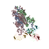
Deposited unit
A: Spike protein S1
B: Repebody (A6)
hetero molecules


Theoretical massNumber of molelcules
Total (without water)52,6253
Polymers52,4042
Non-polymers2211
Water2,144119
1


  • Idetical with deposited unit
  • defined by author
  • Evidence: electron microscopy
TypeNameSymmetry operationNumber
identity operation1_555x,y,z1
Buried area2490 Å2
ΔGint2 kcal/mol
Surface area20560 Å2
Unit cell
Length a, b, c (Å)135.966, 135.966, 69.556
Angle α, β, γ (deg.)90.000, 90.000, 90.000
Int Tables number80
Space group name H-MI41
Space group name HallI4bw
Symmetry operation#1: x,y,z
#2: -y+1/2,x,z+3/4
#3: y+1/2,-x,z+3/4
#4: -x,-y,z
#5: x+1/2,y+1/2,z+1/2
#6: -y+1,x+1/2,z+5/4
#7: y+1,-x+1/2,z+5/4
#8: -x+1/2,-y+1/2,z+1/2

-
Components

#1: Protein Spike protein S1


Mass: 22218.936 Da / Num. of mol.: 1
Source method: isolated from a genetically manipulated source
Source: (gene. exp.) Severe acute respiratory syndrome coronavirus 2
Gene: S, 2 / Production host: Spodoptera frugiperda (fall armyworm) / References: UniProt: P0DTC2
#2: Protein Repebody (A6)


Mass: 30184.621 Da / Num. of mol.: 1
Source method: isolated from a genetically manipulated source
Source: (gene. exp.) Cyclostomatida (invertebrata) / Production host: Escherichia coli BL21(DE3) (bacteria)
#3: Sugar ChemComp-NAG / 2-acetamido-2-deoxy-beta-D-glucopyranose / N-acetyl-beta-D-glucosamine / 2-acetamido-2-deoxy-beta-D-glucose / 2-acetamido-2-deoxy-D-glucose / 2-acetamido-2-deoxy-glucose / N-ACETYL-D-GLUCOSAMINE


Type: D-saccharide, beta linking / Mass: 221.208 Da / Num. of mol.: 1 / Source method: obtained synthetically / Formula: C8H15NO6 / Feature type: SUBJECT OF INVESTIGATION
IdentifierTypeProgram
DGlcpNAcbCONDENSED IUPAC CARBOHYDRATE SYMBOLGMML 1.0
N-acetyl-b-D-glucopyranosamineCOMMON NAMEGMML 1.0
b-D-GlcpNAcIUPAC CARBOHYDRATE SYMBOLPDB-CARE 1.0
GlcNAcSNFG CARBOHYDRATE SYMBOLGMML 1.0
#4: Water ChemComp-HOH / water


Mass: 18.015 Da / Num. of mol.: 119 / Source method: isolated from a natural source / Formula: H2O
Has ligand of interestY
Has protein modificationY

-
Experimental details

-
Experiment

ExperimentMethod: X-RAY DIFFRACTION / Number of used crystals: 1

-
Sample preparation

CrystalDensity Matthews: 3.14 Å3/Da / Density % sol: 60.89 %
Crystal growTemperature: 293 K / Method: vapor diffusion, sitting drop
Details: 0.2 M sodium citrate tribasic dihydrate, 20 % (w/v) PEG 3350

-
Data collection

DiffractionMean temperature: 100 K / Serial crystal experiment: N
Diffraction sourceSource: SYNCHROTRON / Site: PAL/PLS / Beamline: 5C (4A) / Wavelength: 1 Å
DetectorType: DECTRIS EIGER X 9M / Detector: PIXEL / Date: Feb 19, 2022
RadiationProtocol: SINGLE WAVELENGTH / Monochromatic (M) / Laue (L): M / Scattering type: x-ray
Radiation wavelengthWavelength: 1 Å / Relative weight: 1
ReflectionResolution: 1.96→29.8 Å / Num. obs: 45636 / % possible obs: 99.93 % / Redundancy: 13.5 % / Biso Wilson estimate: 40.63 Å2 / CC1/2: 0.999 / CC star: 1 / Rmerge(I) obs: 0.08468 / Net I/σ(I): 19.88
Reflection shellResolution: 1.96→2.03 Å / Redundancy: 14.2 % / Rmerge(I) obs: 1.972 / Mean I/σ(I) obs: 1.52 / Num. unique obs: 4541 / CC1/2: 0.744 / CC star: 0.924 / % possible all: 99.98

-
Processing

Software
NameVersionClassification
PHENIX1.19.2_4158refinement
XDSdata reduction
XDSdata scaling
PHASERphasing
RefinementMethod to determine structure: SAD / Resolution: 1.96→29.8 Å / SU ML: 0.2715 / Cross valid method: FREE R-VALUE / σ(F): 1.35 / Phase error: 36.1058
Stereochemistry target values: GeoStd + Monomer Library + CDL v1.2
RfactorNum. reflection% reflection
Rfree0.2585 2734 3.06 %
Rwork0.2355 86628 -
obs0.2362 45620 99.97 %
Solvent computationShrinkage radii: 0.9 Å / VDW probe radii: 1.11 Å / Solvent model: FLAT BULK SOLVENT MODEL
Displacement parametersBiso mean: 53.74 Å2
Refinement stepCycle: LAST / Resolution: 1.96→29.8 Å
ProteinNucleic acidLigandSolventTotal
Num. atoms3624 0 14 119 3757
Refine LS restraints
Refine-IDTypeDev idealNumber
X-RAY DIFFRACTIONf_bond_d0.00823724
X-RAY DIFFRACTIONf_angle_d1.08035066
X-RAY DIFFRACTIONf_chiral_restr0.0654571
X-RAY DIFFRACTIONf_plane_restr0.0061653
X-RAY DIFFRACTIONf_dihedral_angle_d13.51191367
LS refinement shell
Resolution (Å)Rfactor RfreeNum. reflection RfreeRfactor RworkNum. reflection RworkRefine-ID% reflection obs (%)
1.96-1.990.43311380.46974308X-RAY DIFFRACTION99.93
1.99-2.030.38031380.41814358X-RAY DIFFRACTION99.96
2.03-2.070.4241280.39644307X-RAY DIFFRACTION99.98
2.07-2.110.34741420.38224390X-RAY DIFFRACTION99.98
2.11-2.160.37841400.3774285X-RAY DIFFRACTION99.89
2.16-2.210.45551360.35734303X-RAY DIFFRACTION99.98
2.21-2.260.34461370.29734358X-RAY DIFFRACTION99.96
2.26-2.320.28951380.28484372X-RAY DIFFRACTION99.91
2.32-2.390.33821380.26844296X-RAY DIFFRACTION100
2.39-2.470.32241360.26364338X-RAY DIFFRACTION99.98
2.47-2.560.31241320.27234338X-RAY DIFFRACTION100
2.56-2.660.33261330.27124315X-RAY DIFFRACTION100
2.66-2.780.28941420.26164345X-RAY DIFFRACTION99.96
2.78-2.930.32291360.28654329X-RAY DIFFRACTION99.96
2.93-3.110.3171340.26744324X-RAY DIFFRACTION100
3.11-3.350.25361440.24144317X-RAY DIFFRACTION99.96
3.35-3.690.22481360.21374348X-RAY DIFFRACTION100
3.69-4.220.20921360.19444320X-RAY DIFFRACTION100
4.22-5.310.20161360.16814340X-RAY DIFFRACTION100
5.31-29.80.19521340.18114337X-RAY DIFFRACTION100

+
About Yorodumi

-
News

-
Feb 9, 2022. New format data for meta-information of EMDB entries

New format data for meta-information of EMDB entries

  • Version 3 of the EMDB header file is now the official format.
  • The previous official version 1.9 will be removed from the archive.

Related info.:EMDB header

External links:wwPDB to switch to version 3 of the EMDB data model

-
Aug 12, 2020. Covid-19 info

Covid-19 info

URL: https://pdbj.org/emnavi/covid19.php

New page: Covid-19 featured information page in EM Navigator.

Related info.:Covid-19 info / Mar 5, 2020. Novel coronavirus structure data

+
Mar 5, 2020. Novel coronavirus structure data

Novel coronavirus structure data

Related info.:Yorodumi Speices / Aug 12, 2020. Covid-19 info

External links:COVID-19 featured content - PDBj / Molecule of the Month (242):Coronavirus Proteases

+
Jan 31, 2019. EMDB accession codes are about to change! (news from PDBe EMDB page)

EMDB accession codes are about to change! (news from PDBe EMDB page)

  • The allocation of 4 digits for EMDB accession codes will soon come to an end. Whilst these codes will remain in use, new EMDB accession codes will include an additional digit and will expand incrementally as the available range of codes is exhausted. The current 4-digit format prefixed with “EMD-” (i.e. EMD-XXXX) will advance to a 5-digit format (i.e. EMD-XXXXX), and so on. It is currently estimated that the 4-digit codes will be depleted around Spring 2019, at which point the 5-digit format will come into force.
  • The EM Navigator/Yorodumi systems omit the EMD- prefix.

Related info.:Q: What is EMD? / ID/Accession-code notation in Yorodumi/EM Navigator

External links:EMDB Accession Codes are Changing Soon! / Contact to PDBj

+
Jul 12, 2017. Major update of PDB

Major update of PDB

  • wwPDB released updated PDB data conforming to the new PDBx/mmCIF dictionary.
  • This is a major update changing the version number from 4 to 5, and with Remediation, in which all the entries are updated.
  • In this update, many items about electron microscopy experimental information are reorganized (e.g. em_software).
  • Now, EM Navigator and Yorodumi are based on the updated data.

External links:wwPDB Remediation / Enriched Model Files Conforming to OneDep Data Standards Now Available in the PDB FTP Archive

-
Yorodumi

Thousand views of thousand structures

  • Yorodumi is a browser for structure data from EMDB, PDB, SASBDB, etc.
  • This page is also the successor to EM Navigator detail page, and also detail information page/front-end page for Omokage search.
  • The word "yorodu" (or yorozu) is an old Japanese word meaning "ten thousand". "mi" (miru) is to see.

Related info.:EMDB / PDB / SASBDB / Comparison of 3 databanks / Yorodumi Search / Aug 31, 2016. New EM Navigator & Yorodumi / Yorodumi Papers / Jmol/JSmol / Function and homology information / Changes in new EM Navigator and Yorodumi

Read more