[English] 日本語
Yorodumi
- PDB-7ycl: Crystal structure of SARS-CoV-2 Spike RBD in complex with IS-9A Fab -

+
Open data


ID or keywords:

Loading...

-
Basic information

Entry
Database: PDB / ID: 7ycl
TitleCrystal structure of SARS-CoV-2 Spike RBD in complex with IS-9A Fab
Components
  • IS-9A Fab heavy chain
  • IS-9A Fab light chain
  • Spike protein S1
KeywordsVIRAL PROTEIN/IMMUNE SYSTEM / antibody / SARS-CoV-2 / Spike / RBD / class 4 / VIRAL PROTEIN-IMMUNE SYSTEM complex
Function / homology
Function and homology information


Maturation of spike protein / viral translation / Translation of Structural Proteins / Virion Assembly and Release / host cell surface / host extracellular space / suppression by virus of host tetherin activity / Induction of Cell-Cell Fusion / structural constituent of virion / host cell endoplasmic reticulum-Golgi intermediate compartment membrane ...Maturation of spike protein / viral translation / Translation of Structural Proteins / Virion Assembly and Release / host cell surface / host extracellular space / suppression by virus of host tetherin activity / Induction of Cell-Cell Fusion / structural constituent of virion / host cell endoplasmic reticulum-Golgi intermediate compartment membrane / entry receptor-mediated virion attachment to host cell / receptor-mediated endocytosis of virus by host cell / Attachment and Entry / membrane fusion / positive regulation of viral entry into host cell / receptor-mediated virion attachment to host cell / receptor ligand activity / host cell surface receptor binding / fusion of virus membrane with host plasma membrane / fusion of virus membrane with host endosome membrane / viral envelope / symbiont-mediated suppression of host type I interferon-mediated signaling pathway / virion attachment to host cell / SARS-CoV-2 activates/modulates innate and adaptive immune responses / host cell plasma membrane / virion membrane / membrane / identical protein binding / plasma membrane
Similarity search - Function
Spike (S) protein S1 subunit, receptor-binding domain, SARS-CoV-2 / Spike (S) protein S1 subunit, N-terminal domain, SARS-CoV-like / Betacoronavirus spike (S) glycoprotein S1 subunit N-terminal (NTD) domain profile. / Spike glycoprotein, N-terminal domain superfamily / Betacoronavirus spike (S) glycoprotein S1 subunit C-terminal (CTD) domain profile. / Spike glycoprotein, betacoronavirus / Spike (S) protein S1 subunit, receptor-binding domain, betacoronavirus / Spike S1 subunit, receptor binding domain superfamily, betacoronavirus / Betacoronavirus spike glycoprotein S1, receptor binding / Spike glycoprotein S1, N-terminal domain, betacoronavirus-like ...Spike (S) protein S1 subunit, receptor-binding domain, SARS-CoV-2 / Spike (S) protein S1 subunit, N-terminal domain, SARS-CoV-like / Betacoronavirus spike (S) glycoprotein S1 subunit N-terminal (NTD) domain profile. / Spike glycoprotein, N-terminal domain superfamily / Betacoronavirus spike (S) glycoprotein S1 subunit C-terminal (CTD) domain profile. / Spike glycoprotein, betacoronavirus / Spike (S) protein S1 subunit, receptor-binding domain, betacoronavirus / Spike S1 subunit, receptor binding domain superfamily, betacoronavirus / Betacoronavirus spike glycoprotein S1, receptor binding / Spike glycoprotein S1, N-terminal domain, betacoronavirus-like / Betacoronavirus-like spike glycoprotein S1, N-terminal / Spike glycoprotein S2, coronavirus, heptad repeat 1 / Spike glycoprotein S2, coronavirus, heptad repeat 2 / Coronavirus spike (S) glycoprotein S2 subunit heptad repeat 2 (HR2) region profile. / Coronavirus spike (S) glycoprotein S2 subunit heptad repeat 1 (HR1) region profile. / Spike glycoprotein S2 superfamily, coronavirus / Spike glycoprotein S2, coronavirus / Coronavirus spike glycoprotein S2 / Coronavirus spike glycoprotein S1, C-terminal / Coronavirus spike glycoprotein S1, C-terminal
Similarity search - Domain/homology
Biological speciesHomo sapiens (human)
Severe acute respiratory syndrome coronavirus 2
MethodX-RAY DIFFRACTION / SYNCHROTRON / MOLECULAR REPLACEMENT / Resolution: 2.13 Å
AuthorsMohapatra, A. / Chen, X.
Funding support Taiwan, 1items
OrganizationGrant numberCountry
Ministry of Science and Technology (MoST, Taiwan) Taiwan
CitationJournal: Nat Commun / Year: 2023
Title: Structural basis for a conserved neutralization epitope on the receptor-binding domain of SARS-CoV-2.
Authors: Kuan-Ying A Huang / Xiaorui Chen / Arpita Mohapatra / Hong Thuy Vy Nguyen / Lisa Schimanski / Tiong Kit Tan / Pramila Rijal / Susan K Vester / Rory A Hills / Mark Howarth / Jennifer R Keeffe ...Authors: Kuan-Ying A Huang / Xiaorui Chen / Arpita Mohapatra / Hong Thuy Vy Nguyen / Lisa Schimanski / Tiong Kit Tan / Pramila Rijal / Susan K Vester / Rory A Hills / Mark Howarth / Jennifer R Keeffe / Alexander A Cohen / Leesa M Kakutani / Yi-Min Wu / Md Shahed-Al-Mahmud / Yu-Chi Chou / Pamela J Bjorkman / Alain R Townsend / Che Ma /
Abstract: Antibody-mediated immunity plays a crucial role in protection against SARS-CoV-2 infection. We isolated a panel of neutralizing anti-receptor-binding domain (RBD) antibodies elicited upon natural ...Antibody-mediated immunity plays a crucial role in protection against SARS-CoV-2 infection. We isolated a panel of neutralizing anti-receptor-binding domain (RBD) antibodies elicited upon natural infection and vaccination and showed that they recognize an immunogenic patch on the internal surface of the core RBD, which faces inwards and is hidden in the "down" state. These antibodies broadly neutralize wild type (Wuhan-Hu-1) SARS-CoV-2, Beta and Delta variants and some are effective against other sarbecoviruses. We observed a continuum of partially overlapping antibody epitopes from lower to upper part of the inner face of the RBD and some antibodies extend towards the receptor-binding motif. The majority of antibodies are substantially compromised by three mutational hotspots (S371L/F, S373P and S375F) in the lower part of the Omicron BA.1, BA.2 and BA.4/5 RBD. By contrast, antibody IY-2A induces a partial unfolding of this variable region and interacts with a conserved conformational epitope to tolerate all antigenic variations and neutralize diverse sarbecoviruses as well. This finding establishes that antibody recognition is not limited to the normal surface structures on the RBD. In conclusion, the delineation of functionally and structurally conserved RBD epitopes highlights potential vaccine and therapeutic candidates for COVID-19.
History
DepositionJul 1, 2022Deposition site: PDBJ / Processing site: PDBJ
Revision 1.0Feb 1, 2023Provider: repository / Type: Initial release
Revision 1.1Nov 29, 2023Group: Data collection / Refinement description
Category: chem_comp_atom / chem_comp_bond / pdbx_initial_refinement_model

-
Structure visualization

Structure viewerMolecule:
MolmilJmol/JSmol

Downloads & links

-
Assembly

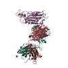
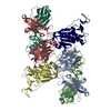
Deposited unit
A: IS-9A Fab heavy chain
B: IS-9A Fab light chain
R: Spike protein S1
L: IS-9A Fab light chain
H: IS-9A Fab heavy chain
D: Spike protein S1
hetero molecules


Theoretical massNumber of molelcules
Total (without water)141,9247
Polymers141,3546
Non-polymers5711
Water19,8351101
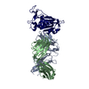
1
A: IS-9A Fab heavy chain
B: IS-9A Fab light chain
R: Spike protein S1


Theoretical massNumber of molelcules
Total (without water)70,6773
Polymers70,6773
Non-polymers00
Water543
TypeNameSymmetry operationNumber
identity operation1_555x,y,z1
2
L: IS-9A Fab light chain
H: IS-9A Fab heavy chain
D: Spike protein S1
hetero molecules


Theoretical massNumber of molelcules
Total (without water)71,2474
Polymers70,6773
Non-polymers5711
Water543
TypeNameSymmetry operationNumber
identity operation1_555x,y,z1
Unit cell
Length a, b, c (Å)85.279, 86.860, 98.089
Angle α, β, γ (deg.)90.000, 108.660, 90.000
Int Tables number4
Space group name H-MP1211
Noncrystallographic symmetry (NCS)NCS domain:
IDEns-IDDetails
11(chain A and (resid 1 through 135 or resid 143 through 224))
21chain H
12(chain B and (resid 1 through 141 or resid 143 through 213))
22(chain L and (resid 1 through 141 or resid 143 through 213))
13(chain D and (resid 334 through 465 or resid 467 through 527))
23(chain R and (resid 334 through 465 or resid 467 through 527))

NCS domain segments:
Dom-IDComponent-IDEns-IDSelection detailsAuth asym-IDAuth seq-ID
111(chain A and (resid 1 through 135 or resid 143 through 224))A1 - 135
121(chain A and (resid 1 through 135 or resid 143 through 224))A143 - 224
211chain HH1 - 215
112(chain B and (resid 1 through 141 or resid 143 through 213))B1 - 141
122(chain B and (resid 1 through 141 or resid 143 through 213))B143 - 213
212(chain L and (resid 1 through 141 or resid 143 through 213))L1 - 141
222(chain L and (resid 1 through 141 or resid 143 through 213))L143 - 213
113(chain D and (resid 334 through 465 or resid 467 through 527))D334 - 465
123(chain D and (resid 334 through 465 or resid 467 through 527))D467 - 527
213(chain R and (resid 334 through 465 or resid 467 through 527))R334 - 465
223(chain R and (resid 334 through 465 or resid 467 through 527))R467 - 527

NCS ensembles :
ID
1
2
3

-
Components

#1: Antibody IS-9A Fab heavy chain


Mass: 24225.162 Da / Num. of mol.: 2
Source method: isolated from a genetically manipulated source
Source: (gene. exp.) Homo sapiens (human) / Production host: Homo sapiens (human)
#2: Antibody IS-9A Fab light chain


Mass: 23403.809 Da / Num. of mol.: 2
Source method: isolated from a genetically manipulated source
Source: (gene. exp.) Homo sapiens (human) / Production host: Homo sapiens (human)
#3: Protein Spike protein S1


Mass: 23047.816 Da / Num. of mol.: 2
Source method: isolated from a genetically manipulated source
Source: (gene. exp.) Severe acute respiratory syndrome coronavirus 2
Gene: S, 2 / Production host: Homo sapiens (human) / References: UniProt: P0DTC2
#4: Polysaccharide 2-acetamido-2-deoxy-beta-D-glucopyranose-(1-4)-[alpha-L-fucopyranose-(1-6)]2-acetamido-2-deoxy-beta- ...2-acetamido-2-deoxy-beta-D-glucopyranose-(1-4)-[alpha-L-fucopyranose-(1-6)]2-acetamido-2-deoxy-beta-D-glucopyranose


Type: oligosaccharide / Mass: 570.542 Da / Num. of mol.: 1
Source method: isolated from a genetically manipulated source
DescriptorTypeProgram
DGlcpNAcb1-4[LFucpa1-6]DGlcpNAcb1-ROHGlycam Condensed SequenceGMML 1.0
WURCS=2.0/2,3,2/[a2122h-1b_1-5_2*NCC/3=O][a1221m-1a_1-5]/1-1-2/a4-b1_a6-c1WURCSPDB2Glycan 1.1.0
[][D-1-deoxy-GlcpNAc]{[(4+1)][b-D-GlcpNAc]{}[(6+1)][a-L-Fucp]{}}LINUCSPDB-CARE
#5: Water ChemComp-HOH / water / Water


Mass: 18.015 Da / Num. of mol.: 1101 / Source method: isolated from a natural source / Formula: H2O
Has ligand of interestN

-
Experimental details

-
Experiment

ExperimentMethod: X-RAY DIFFRACTION / Number of used crystals: 1

-
Sample preparation

CrystalDensity Matthews: 2.34 Å3/Da / Density % sol: 47.34 %
Crystal growTemperature: 293.15 K / Method: vapor diffusion, hanging drop / pH: 8
Details: 2% v/v 1,4-Dioxane, 0.1M Tris pH 8.0, 15% w/v Polyethylene glycol 3350

-
Data collection

DiffractionMean temperature: 100 K / Serial crystal experiment: N
Diffraction sourceSource: SYNCHROTRON / Site: NSRRC / Beamline: BL15A1 / Wavelength: 1 Å
DetectorType: RAYONIX MX300HE / Detector: CCD / Date: Nov 25, 2021
RadiationProtocol: SINGLE WAVELENGTH / Monochromatic (M) / Laue (L): M / Scattering type: x-ray
Radiation wavelengthWavelength: 1 Å / Relative weight: 1
ReflectionResolution: 2.13→33.95 Å / Num. obs: 75756 / % possible obs: 99.7 % / Redundancy: 3.7 % / Biso Wilson estimate: 22.9 Å2 / CC1/2: 0.997 / Rmerge(I) obs: 0.084 / Rpim(I) all: 0.05 / Rrim(I) all: 0.098 / Net I/σ(I): 9.7 / Num. measured all: 283773 / Scaling rejects: 42
Reflection shell

Diffraction-ID: 1

Resolution (Å)Redundancy (%)Rmerge(I) obsNum. measured allNum. unique obsCC1/2Rpim(I) allRrim(I) allNet I/σ(I) obs% possible all
2.13-2.1730.3191310943270.8680.2210.3913.396.6
10.65-33.953.50.03321676270.9980.0210.03919.297

-
Processing

Software
NameVersionClassification
PHENIX1.19.2_4158refinement
Aimless0.7.4data scaling
PDB_EXTRACT3.27data extraction
MOSFLMdata reduction
PHASERphasing
RefinementMethod to determine structure: MOLECULAR REPLACEMENT
Starting model: 6ZER
Resolution: 2.13→32.62 Å / SU ML: 0.22 / Cross valid method: THROUGHOUT / σ(F): 1.36 / Phase error: 22.73 / Stereochemistry target values: ML
RfactorNum. reflection% reflection
Rfree0.2175 3737 4.94 %
Rwork0.1735 71946 -
obs0.1756 75683 99.7 %
Solvent computationShrinkage radii: 0.9 Å / VDW probe radii: 1.11 Å / Solvent model: FLAT BULK SOLVENT MODEL
Displacement parametersBiso max: 75.31 Å2 / Biso mean: 27.6311 Å2 / Biso min: 5.35 Å2
Refinement stepCycle: final / Resolution: 2.13→32.62 Å
ProteinNucleic acidLigandSolventTotal
Num. atoms9676 0 38 1101 10815
Biso mean--61.08 33.54 -
Num. residues----1252
Refine LS restraints NCS
Ens-IDDom-IDAuth asym-IDNumberRefine-IDRmsType
11A1947X-RAY DIFFRACTION12.525TORSIONAL
12H1947X-RAY DIFFRACTION12.525TORSIONAL
21B1918X-RAY DIFFRACTION12.525TORSIONAL
22L1918X-RAY DIFFRACTION12.525TORSIONAL
31D1760X-RAY DIFFRACTION12.525TORSIONAL
32R1760X-RAY DIFFRACTION12.525TORSIONAL
LS refinement shell

Refine-ID: X-RAY DIFFRACTION / Rfactor Rfree error: 0 / Total num. of bins used: 27

Resolution (Å)Rfactor RfreeNum. reflection RfreeRfactor RworkNum. reflection RworkNum. reflection all% reflection obs (%)
2.13-2.160.27181370.22952486262396
2.16-2.190.26811280.22482649277798
2.19-2.220.31551510.223626272778100
2.22-2.250.29641450.22526512796100
2.25-2.280.25181360.223126592795100
2.28-2.320.28231430.209826302773100
2.32-2.350.27881410.201526592800100
2.35-2.390.23621580.196426622820100
2.39-2.440.24731310.196326732804100
2.44-2.490.27221470.191526502797100
2.49-2.540.26211320.190526592791100
2.54-2.590.25311440.183526642808100
2.59-2.650.24081280.185527052833100
2.65-2.720.23821370.179326492786100
2.72-2.790.2511410.182626602801100
2.79-2.870.2551430.18326442787100
2.87-2.970.25031350.186326872822100
2.97-3.070.23761570.18426642821100
3.07-3.190.22511170.183726812798100
3.19-3.340.19921340.168626632797100
3.34-3.520.23491240.16326972821100
3.52-3.740.18051460.159626712817100
3.74-4.020.18631320.151126952827100
4.02-4.430.14121180.13227012819100
4.43-5.070.14061410.127927132854100
5.07-6.370.18711310.152627062837100
6.38-32.620.18151600.166527412901100

+
About Yorodumi

-
News

-
Feb 9, 2022. New format data for meta-information of EMDB entries

New format data for meta-information of EMDB entries

  • Version 3 of the EMDB header file is now the official format.
  • The previous official version 1.9 will be removed from the archive.

Related info.:EMDB header

External links:wwPDB to switch to version 3 of the EMDB data model

-
Aug 12, 2020. Covid-19 info

Covid-19 info

URL: https://pdbj.org/emnavi/covid19.php

New page: Covid-19 featured information page in EM Navigator.

Related info.:Covid-19 info / Mar 5, 2020. Novel coronavirus structure data

+
Mar 5, 2020. Novel coronavirus structure data

Novel coronavirus structure data

Related info.:Yorodumi Speices / Aug 12, 2020. Covid-19 info

External links:COVID-19 featured content - PDBj / Molecule of the Month (242):Coronavirus Proteases

+
Jan 31, 2019. EMDB accession codes are about to change! (news from PDBe EMDB page)

EMDB accession codes are about to change! (news from PDBe EMDB page)

  • The allocation of 4 digits for EMDB accession codes will soon come to an end. Whilst these codes will remain in use, new EMDB accession codes will include an additional digit and will expand incrementally as the available range of codes is exhausted. The current 4-digit format prefixed with “EMD-” (i.e. EMD-XXXX) will advance to a 5-digit format (i.e. EMD-XXXXX), and so on. It is currently estimated that the 4-digit codes will be depleted around Spring 2019, at which point the 5-digit format will come into force.
  • The EM Navigator/Yorodumi systems omit the EMD- prefix.

Related info.:Q: What is EMD? / ID/Accession-code notation in Yorodumi/EM Navigator

External links:EMDB Accession Codes are Changing Soon! / Contact to PDBj

+
Jul 12, 2017. Major update of PDB

Major update of PDB

  • wwPDB released updated PDB data conforming to the new PDBx/mmCIF dictionary.
  • This is a major update changing the version number from 4 to 5, and with Remediation, in which all the entries are updated.
  • In this update, many items about electron microscopy experimental information are reorganized (e.g. em_software).
  • Now, EM Navigator and Yorodumi are based on the updated data.

External links:wwPDB Remediation / Enriched Model Files Conforming to OneDep Data Standards Now Available in the PDB FTP Archive

-
Yorodumi

Thousand views of thousand structures

  • Yorodumi is a browser for structure data from EMDB, PDB, SASBDB, etc.
  • This page is also the successor to EM Navigator detail page, and also detail information page/front-end page for Omokage search.
  • The word "yorodu" (or yorozu) is an old Japanese word meaning "ten thousand". "mi" (miru) is to see.

Related info.:EMDB / PDB / SASBDB / Comparison of 3 databanks / Yorodumi Search / Aug 31, 2016. New EM Navigator & Yorodumi / Yorodumi Papers / Jmol/JSmol / Function and homology information / Changes in new EM Navigator and Yorodumi

Read more