 Movie
Movie Controller


+ Open data

- Basic information
| Entry | Database: PDB / ID: 7yan | ||||||
|---|---|---|---|---|---|---|---|
| Title | UDP-glucuronosyltransferase2B17 C-terminal domain | ||||||
 Components Components | UDP-glucuronosyltransferase 2B17 | ||||||
 Keywords Keywords | TRANSFERASE / Complex / truncation | ||||||
| Function / homology |  Function and homology information Function and homology information: / glucuronosyltransferase / Glucuronidation / glucuronosyltransferase activity / estrogen metabolic process / Prednisone ADME / Aspirin ADME / steroid metabolic process / xenobiotic metabolic process / endoplasmic reticulum membrane / membrane Similarity search - Function | ||||||
| Biological species |  Homo sapiens (human) Homo sapiens (human) | ||||||
| Method |  X-RAY DIFFRACTION / MOLECULAR REPLACEMENT / Resolution: 2.5 Å X-RAY DIFFRACTION / MOLECULAR REPLACEMENT / Resolution: 2.5 Å | ||||||
 Authors Authors | Wang, C.Y. / Zhang, L. | ||||||
| Funding support |  China, 1items China, 1items
| ||||||
 Citation Citation | Journal: To Be Published Title: UDP-glucuronosyltransferase2B17 C-terminal domain Authors: Wang, C.Y. / Zhang, L. | ||||||
| History |
|
- Structure visualization
| Structure viewer | Molecule: MolmilJmol/JSmol |
|---|
- Downloads & links
-Download
| PDBx/mmCIF format | 7yan.cif.gz | 142 KB | Display | PDBx/mmCIF format |
|---|---|---|---|---|
| PDB format | pdb7yan.ent.gz | 106.4 KB | Display | PDB format |
| PDBx/mmJSON format | 7yan.json.gz | Tree view | PDBx/mmJSON format | |
| Others |  Other downloads Other downloads |
-Validation report
| Summary document | 7yan_validation.pdf.gz | 1.7 MB | Display | wwPDB validaton report |
|---|---|---|---|---|
| Full document | 7yan_full_validation.pdf.gz | 1.7 MB | Display | |
| Data in XML | 7yan_validation.xml.gz | 26.5 KB | Display | |
| Data in CIF | 7yan_validation.cif.gz | 35.1 KB | Display | |
| Arichive directory | https://data.pdbj.org/pub/pdb/validation_reports/ya/7yanftp://data.pdbj.org/pub/pdb/validation_reports/ya/7yan | HTTPS FTP |
-Related structure data
| Related structure data | 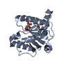 6ipbS S: Starting model for refinement |
|---|---|
| Similar structure data |
-Links
 PDBj
PDBj
- Assembly
| Deposited unit | 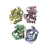
| ||||||||
|---|---|---|---|---|---|---|---|---|---|
| 1 | 
| ||||||||
| 2 | 
| ||||||||
| Unit cell |
|
-Components
| #1: Protein | Mass: 21046.270 Da / Num. of mol.: 4 Source method: isolated from a genetically manipulated source Source: (gene. exp.) Homo sapiens (human) / Gene: UGT2B17 / Production host:  #2: Chemical | ChemComp-TLA /   Mass: 150.087 Da / Num. of mol.: 4 / Source method: obtained synthetically / Formula: C4H6O6 / Feature type: SUBJECT OF INVESTIGATION Mass: 150.087 Da / Num. of mol.: 4 / Source method: obtained synthetically / Formula: C4H6O6 / Feature type: SUBJECT OF INVESTIGATION#3: Water | ChemComp-HOH / |  Mass: 18.015 Da / Num. of mol.: 46 / Source method: isolated from a natural source / Formula: H2O Mass: 18.015 Da / Num. of mol.: 46 / Source method: isolated from a natural source / Formula: H2OHas ligand of interest | Y | |
|---|
-Experimental details
-Experiment
| Experiment | Method: X-RAY DIFFRACTION / Number of used crystals: 1 |
|---|
- Sample preparation
| Crystal | Density Matthews: 1.89 Å3/Da / Density % sol: 34.99 % |
|---|---|
| Crystal grow | Temperature: 298 K / Method: vapor diffusion, sitting drop Details: 20% PEG 6000, 0.2M Na-Tartrate, 0.2M KCl, 2% glycerol |
-Data collection
| Diffraction | Mean temperature: 100 K / Serial crystal experiment: N |
|---|---|
| Diffraction source | Source: ROTATING ANODE / Type: RIGAKU MICROMAX-007 / Wavelength: 1.54184 Å |
| Detector | Type: DECTRIS PILATUS 200K / Detector: PIXEL / Date: Jun 17, 2021 |
| Radiation | Protocol: SINGLE WAVELENGTH / Monochromatic (M) / Laue (L): M / Scattering type: x-ray |
| Radiation wavelength | Wavelength: 1.54184 Å / Relative weight: 1 |
| Reflection | Resolution: 2.5→19.11 Å / Num. obs: 20837 / % possible obs: 97.45 % / Redundancy: 2.7 % / CC1/2: 0.985 / Rmerge(I) obs: 0.0978 / Net I/σ(I): 8.05 |
| Reflection shell | Resolution: 2.5→2.589 Å / Num. unique obs: 2019 / CC1/2: 0.893 |
- Processing
| Software |
| |||||||||||||||||||||||||||||||||||||||||||||||||||||||||||||||||||||||||||||||||||||||||||||||||||||||||||||||||||||||||||||||||||||||||||||||||||||||||||
|---|---|---|---|---|---|---|---|---|---|---|---|---|---|---|---|---|---|---|---|---|---|---|---|---|---|---|---|---|---|---|---|---|---|---|---|---|---|---|---|---|---|---|---|---|---|---|---|---|---|---|---|---|---|---|---|---|---|---|---|---|---|---|---|---|---|---|---|---|---|---|---|---|---|---|---|---|---|---|---|---|---|---|---|---|---|---|---|---|---|---|---|---|---|---|---|---|---|---|---|---|---|---|---|---|---|---|---|---|---|---|---|---|---|---|---|---|---|---|---|---|---|---|---|---|---|---|---|---|---|---|---|---|---|---|---|---|---|---|---|---|---|---|---|---|---|---|---|---|---|---|---|---|---|---|---|---|
| Refinement | Method to determine structure: MOLECULAR REPLACEMENT Starting model: 6IPB Resolution: 2.5→19.11 Å / Cor.coef. Fo:Fc: 0.838 / Cor.coef. Fo:Fc free: 0.763 / SU B: 17.612 / SU ML: 0.388 / Cross valid method: THROUGHOUT / ESU R Free: 0.432 Details: Hydrogens have been added in their riding positions
| |||||||||||||||||||||||||||||||||||||||||||||||||||||||||||||||||||||||||||||||||||||||||||||||||||||||||||||||||||||||||||||||||||||||||||||||||||||||||||
| Solvent computation | Ion probe radii: 0.8 Å / Shrinkage radii: 0.8 Å / VDW probe radii: 1.2 Å / Solvent model: MASK BULK SOLVENT | |||||||||||||||||||||||||||||||||||||||||||||||||||||||||||||||||||||||||||||||||||||||||||||||||||||||||||||||||||||||||||||||||||||||||||||||||||||||||||
| Displacement parameters | Biso mean: 18.094 Å2
| |||||||||||||||||||||||||||||||||||||||||||||||||||||||||||||||||||||||||||||||||||||||||||||||||||||||||||||||||||||||||||||||||||||||||||||||||||||||||||
| Refinement step | Cycle: LAST / Resolution: 2.5→19.11 Å
| |||||||||||||||||||||||||||||||||||||||||||||||||||||||||||||||||||||||||||||||||||||||||||||||||||||||||||||||||||||||||||||||||||||||||||||||||||||||||||
| Refine LS restraints |
| |||||||||||||||||||||||||||||||||||||||||||||||||||||||||||||||||||||||||||||||||||||||||||||||||||||||||||||||||||||||||||||||||||||||||||||||||||||||||||
| LS refinement shell |
|
