 Movie
Movie Controller


+ Open data

- Basic information
| Entry | Database: PDB / ID: 7yam | |||||||||||||||||||||||||||||||||||||||
|---|---|---|---|---|---|---|---|---|---|---|---|---|---|---|---|---|---|---|---|---|---|---|---|---|---|---|---|---|---|---|---|---|---|---|---|---|---|---|---|---|
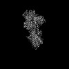
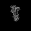
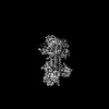
| Title | CryoEM structure of SPCA1a in E2P state | |||||||||||||||||||||||||||||||||||||||
 Components Components | Calcium-transporting ATPase type 2C member 1 | |||||||||||||||||||||||||||||||||||||||
 Keywords Keywords | TRANSPORT PROTEIN / Golgi Ca2+/Mn2+ transporter | |||||||||||||||||||||||||||||||||||||||
| Function / homology |  Function and homology information Function and homology informationGolgi calcium ion homeostasis / Golgi calcium ion transport / P-type manganese transporter activity / trans-Golgi network membrane organization / manganese ion transport / intracellular manganese ion homeostasis / cis-Golgi network membrane / P-type Ca2+ transporter / P-type calcium transporter activity / calcium-dependent cell-cell adhesion ...Golgi calcium ion homeostasis / Golgi calcium ion transport / P-type manganese transporter activity / trans-Golgi network membrane organization / manganese ion transport / intracellular manganese ion homeostasis / cis-Golgi network membrane / P-type Ca2+ transporter / P-type calcium transporter activity / calcium-dependent cell-cell adhesion / positive regulation of Golgi to plasma membrane protein transport / Golgi cisterna membrane / Ion transport by P-type ATPases / epidermis development / trans-Golgi network / calcium ion transmembrane transport / intracellular calcium ion homeostasis / calcium ion transport / manganese ion binding / actin cytoskeleton organization / positive regulation of canonical NF-kappaB signal transduction / Golgi membrane / calcium ion binding / endoplasmic reticulum / Golgi apparatus / ATP hydrolysis activity / ATP binding / metal ion binding / membrane / plasma membrane Similarity search - Function | |||||||||||||||||||||||||||||||||||||||
| Biological species |  Homo sapiens (human) Homo sapiens (human) | |||||||||||||||||||||||||||||||||||||||
| Method | ELECTRON MICROSCOPY / single particle reconstruction / cryo EM / Resolution: 3.3 Å | |||||||||||||||||||||||||||||||||||||||
 Authors Authors | Chen, Z. / Watanabe, S. / Inaba, K. | |||||||||||||||||||||||||||||||||||||||
| Funding support |  Japan, 2items Japan, 2items
| |||||||||||||||||||||||||||||||||||||||
 Citation Citation | Journal: Sci Adv / Year: 2023 Title: Cryo-EM structures of human SPCA1a reveal the mechanism of Ca/Mn transport into the Golgi apparatus. Authors: Zhenghao Chen / Satoshi Watanabe / Hironori Hashida / Michio Inoue / Yasukazu Daigaku / Masahide Kikkawa / Kenji Inaba / Abstract: Secretory pathway Ca/Mn ATPase 1 (SPCA1) actively transports cytosolic Ca and Mn into the Golgi lumen, playing a crucial role in cellular calcium and manganese homeostasis. Detrimental mutations of ...Secretory pathway Ca/Mn ATPase 1 (SPCA1) actively transports cytosolic Ca and Mn into the Golgi lumen, playing a crucial role in cellular calcium and manganese homeostasis. Detrimental mutations of the gene encoding SPCA1 cause Hailey-Hailey disease. Here, using nanobody/megabody technologies, we determined cryo-electron microscopy structures of human SPCA1a in the ATP and Ca/Mn-bound (E1-ATP) state and the metal-free phosphorylated (E2P) state at 3.1- to 3.3-Å resolutions. The structures revealed that Ca and Mn share the same metal ion-binding pocket with similar but notably different coordination geometries in the transmembrane domain, corresponding to the second Ca-binding site in sarco/endoplasmic reticulum Ca-ATPase (SERCA). In the E1-ATP to E2P transition, SPCA1a undergoes similar domain rearrangements to those of SERCA. Meanwhile, SPCA1a shows larger conformational and positional flexibility of the second and sixth transmembrane helices, possibly explaining its wider metal ion specificity. These structural findings illuminate the unique mechanisms of SPCA1a-mediated Ca/Mn transport. | |||||||||||||||||||||||||||||||||||||||
| History |
|
- Structure visualization
| Structure viewer | Molecule: MolmilJmol/JSmol |
|---|
- Downloads & links
-Download
| PDBx/mmCIF format | 7yam.cif.gz | 161.5 KB | Display | PDBx/mmCIF format |
|---|---|---|---|---|
| PDB format | pdb7yam.ent.gz | 123 KB | Display | PDB format |
| PDBx/mmJSON format | 7yam.json.gz | Tree view | PDBx/mmJSON format | |
| Others |  Other downloads Other downloads |
-Validation report
| Arichive directory | https://data.pdbj.org/pub/pdb/validation_reports/ya/7yamftp://data.pdbj.org/pub/pdb/validation_reports/ya/7yam | HTTPS FTP |
|---|
-Related structure data
| Related structure data | 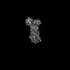 33717MC 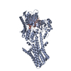 7yagC 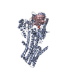 7yahC 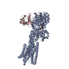 7yaiC 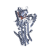 7yajC M: map data used to model this data C: citing same article ( |
|---|---|
| Similar structure data |

-Links
 PDBj
PDBj


- Assembly
| Deposited unit | 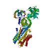
|
|---|---|
| 1 |
|
-Components
| #1: Protein | Mass: 103574.203 Da / Num. of mol.: 1 Source method: isolated from a genetically manipulated source Source: (gene. exp.) Homo sapiens (human) / Gene: ATP2C1, KIAA1347, PMR1L, HUSSY-28 / Production host: Homo sapiens (human) / References: UniProt: P98194, P-type Ca2+ transporter |
|---|---|
| #2: Chemical | ChemComp-BEF /   Mass: 66.007 Da / Num. of mol.: 1 / Source method: obtained synthetically / Formula: BeF3 / Feature type: SUBJECT OF INVESTIGATION Mass: 66.007 Da / Num. of mol.: 1 / Source method: obtained synthetically / Formula: BeF3 / Feature type: SUBJECT OF INVESTIGATION |
| #3: Chemical | ChemComp-MG /   Mass: 24.305 Da / Num. of mol.: 1 / Source method: obtained synthetically / Formula: Mg / Feature type: SUBJECT OF INVESTIGATION Mass: 24.305 Da / Num. of mol.: 1 / Source method: obtained synthetically / Formula: Mg / Feature type: SUBJECT OF INVESTIGATION |
| Has ligand of interest | Y |
| Has protein modification | N |
-Experimental details
-Experiment
| Experiment | Method: ELECTRON MICROSCOPY |
|---|---|
| EM experiment | Aggregation state: PARTICLE / 3D reconstruction method: single particle reconstruction |
- Sample preparation
| Component | Name: SPCA1a in E2P state / Type: COMPLEX / Entity ID: #1 / Source: RECOMBINANT |
|---|---|
| Source (natural) | Organism: Homo sapiens (human) |
| Source (recombinant) | Organism: Homo sapiens (human) |
| Buffer solution | pH: 6 |
| Specimen | Embedding applied: NO / Shadowing applied: NO / Staining applied: NO / Vitrification applied: YES |
| Vitrification | Cryogen name: ETHANE |
- Electron microscopy imaging
| Experimental equipment |  Model: Titan Krios / Image courtesy: FEI Company |
|---|---|
| Microscopy | Model: FEI TITAN KRIOS |
| Electron gun | Electron source:  FIELD EMISSION GUN / Accelerating voltage: 300 kV / Illumination mode: OTHER FIELD EMISSION GUN / Accelerating voltage: 300 kV / Illumination mode: OTHER |
| Electron lens | Mode: BRIGHT FIELD / Nominal defocus max: 1800 nm / Nominal defocus min: 1000 nm |
| Image recording | Electron dose: 49 e/Å2 / Film or detector model: GATAN K3 (6k x 4k) |
- Processing
| Software | Name: PHENIX / Version: 1.19.2_4158: / Classification: refinement | ||||||||||||||||||||||||
|---|---|---|---|---|---|---|---|---|---|---|---|---|---|---|---|---|---|---|---|---|---|---|---|---|---|
| EM software | Name: PHENIX / Category: model refinement | ||||||||||||||||||||||||
| CTF correction | Type: NONE | ||||||||||||||||||||||||
| 3D reconstruction | Resolution: 3.3 Å / Resolution method: FSC 0.143 CUT-OFF / Num. of particles: 260172 / Symmetry type: POINT | ||||||||||||||||||||||||
| Refine LS restraints |
|
