Japan Agency for Medical Research and Development (AMED)
21gm1410006h0001, JP19am0101115
Japan
Japan Society for the Promotion of Science (JSPS)
18H03978, 21H04758, 21H05247, 21K15036
Japan
Citation
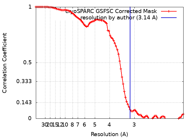
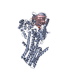
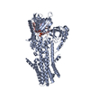
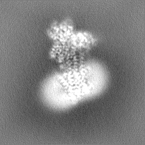
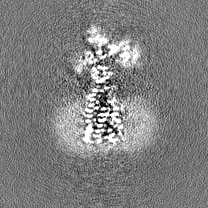
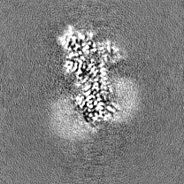
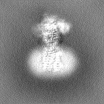
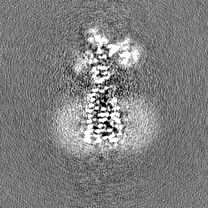
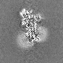
Journal: Sci Adv / Year: 2023 Title: Cryo-EM structures of human SPCA1a reveal the mechanism of Ca/Mn transport into the Golgi apparatus. Authors: Zhenghao Chen / Satoshi Watanabe / Hironori Hashida / Michio Inoue / Yasukazu Daigaku / Masahide Kikkawa / Kenji Inaba / Abstract: Secretory pathway Ca/Mn ATPase 1 (SPCA1) actively transports cytosolic Ca and Mn into the Golgi lumen, playing a crucial role in cellular calcium and manganese homeostasis. Detrimental mutations of ...Secretory pathway Ca/Mn ATPase 1 (SPCA1) actively transports cytosolic Ca and Mn into the Golgi lumen, playing a crucial role in cellular calcium and manganese homeostasis. Detrimental mutations of the gene encoding SPCA1 cause Hailey-Hailey disease. Here, using nanobody/megabody technologies, we determined cryo-electron microscopy structures of human SPCA1a in the ATP and Ca/Mn-bound (E1-ATP) state and the metal-free phosphorylated (E2P) state at 3.1- to 3.3-Å resolutions. The structures revealed that Ca and Mn share the same metal ion-binding pocket with similar but notably different coordination geometries in the transmembrane domain, corresponding to the second Ca-binding site in sarco/endoplasmic reticulum Ca-ATPase (SERCA). In the E1-ATP to E2P transition, SPCA1a undergoes similar domain rearrangements to those of SERCA. Meanwhile, SPCA1a shows larger conformational and positional flexibility of the second and sixth transmembrane helices, possibly explaining its wider metal ion specificity. These structural findings illuminate the unique mechanisms of SPCA1a-mediated Ca/Mn transport.
In the structure databanks used in Yorodumi, some data are registered as the other names, "COVID-19 virus" and "2019-nCoV". Here are the details of the virus and the list of structure data.
Jan 31, 2019. EMDB accession codes are about to change! (news from PDBe EMDB page)
EMDB accession codes are about to change! (news from PDBe EMDB page)
The allocation of 4 digits for EMDB accession codes will soon come to an end. Whilst these codes will remain in use, new EMDB accession codes will include an additional digit and will expand incrementally as the available range of codes is exhausted. The current 4-digit format prefixed with “EMD-” (i.e. EMD-XXXX) will advance to a 5-digit format (i.e. EMD-XXXXX), and so on. It is currently estimated that the 4-digit codes will be depleted around Spring 2019, at which point the 5-digit format will come into force.
The EM Navigator/Yorodumi systems omit the EMD- prefix.
Related info.:Q: What is EMD? / ID/Accession-code notation in Yorodumi/EM Navigator
Yorodumi is a browser for structure data from EMDB, PDB, SASBDB, etc.
This page is also the successor to EM Navigator detail page, and also detail information page/front-end page for Omokage search.
The word "yorodu" (or yorozu) is an old Japanese word meaning "ten thousand". "mi" (miru) is to see.
Related info.:EMDB / PDB / SASBDB / Comparison of 3 databanks / Yorodumi Search / Aug 31, 2016. New EM Navigator & Yorodumi / Yorodumi Papers / Jmol/JSmol / Function and homology information / Changes in new EM Navigator and Yorodumi
 Movie
Movie Controller


 Open data
 Basic information
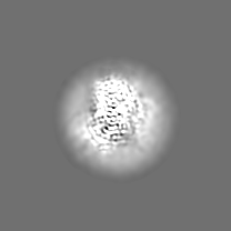
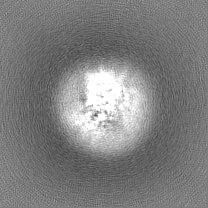
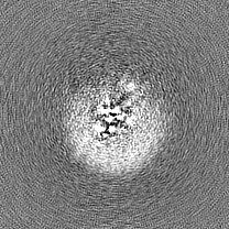
 Map data
 Sample
 Keywords
 Function and homology information
 Homo sapiens (human) /
 Authors
 Japan, 2 items
  Citation
 Structure visualization
 Downloads & links
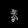
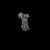
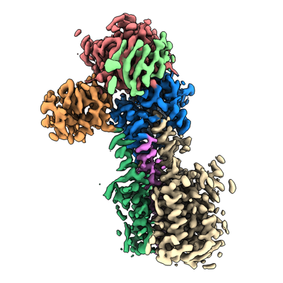 emd_33713.png
 http://ftp.pdbj.org/pub/emdb/structures/EMD-33713


 Z (Sec.)
 Y (Row.)
 X (Col.)


 Sample components

 Processing
 Electron microscopy
 FIELD EMISSION GUN