 Movie
Movie Controller


+ Open data

- Basic information
| Entry | Database: PDB / ID: 7y8c | ||||||
|---|---|---|---|---|---|---|---|
| Title | Crystal structure of CotA laccase complexed with syringaldehyde | ||||||
 Components Components | Spore coat protein A | ||||||
 Keywords Keywords | OXIDOREDUCTASE / laccase | ||||||
| Function / homology |  Function and homology information Function and homology informationbilirubin oxidase / laccase / sporulation resulting in formation of a cellular spore / outer membrane-bounded periplasmic space / oxidoreductase activity / copper ion binding Similarity search - Function | ||||||
| Biological species |  | ||||||
| Method |  X-RAY DIFFRACTION / SYNCHROTRON / MOLECULAR REPLACEMENT / Resolution: 2 Å X-RAY DIFFRACTION / SYNCHROTRON / MOLECULAR REPLACEMENT / Resolution: 2 Å | ||||||
 Authors Authors | Liu, Z.C. / Xie, T. / Wang, G.G. | ||||||
| Funding support | 1items
| ||||||
 Citation Citation | Journal: Int.J.Biol.Macromol. / Year: 2023 Title: Molecular insights into substrate promiscuity of CotA laccase catalyzing lignin-phenol derivatives. Authors: Li, J. / Liu, Z. / Zhao, J. / Wang, G. / Xie, T. | ||||||
| History |
|
- Structure visualization
| Structure viewer | Molecule: MolmilJmol/JSmol |
|---|
- Downloads & links
-Download
| PDBx/mmCIF format | 7y8c.cif.gz | 225.8 KB | Display | PDBx/mmCIF format |
|---|---|---|---|---|
| PDB format | pdb7y8c.ent.gz | 176.9 KB | Display | PDB format |
| PDBx/mmJSON format | 7y8c.json.gz | Tree view | PDBx/mmJSON format | |
| Others |  Other downloads Other downloads |
-Validation report
| Summary document | 7y8c_validation.pdf.gz | 717 KB | Display | wwPDB validaton report |
|---|---|---|---|---|
| Full document | 7y8c_full_validation.pdf.gz | 726.5 KB | Display | |
| Data in XML | 7y8c_validation.xml.gz | 42.6 KB | Display | |
| Data in CIF | 7y8c_validation.cif.gz | 63.1 KB | Display | |
| Arichive directory | https://data.pdbj.org/pub/pdb/validation_reports/y8/7y8cftp://data.pdbj.org/pub/pdb/validation_reports/y8/7y8c | HTTPS FTP |
-Related structure data
| Related structure data |  7y8bC  4q89S C: citing same article ( S: Starting model for refinement |
|---|---|
| Similar structure data |
-Links
 PDBj
PDBj- Assembly
| Deposited unit | 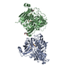
| ||||||||
|---|---|---|---|---|---|---|---|---|---|
| 1 | 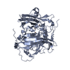
| ||||||||
| 2 | 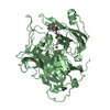
| ||||||||
| Unit cell |
|
-Components
| #1: Protein | Mass: 58574.789 Da / Num. of mol.: 2 Source method: isolated from a genetically manipulated source Source: (gene. exp.) Gene: cotA, pig, BSU06300 / Production host: #2: Chemical | ChemComp-CU /   Mass: 63.546 Da / Num. of mol.: 8 / Source method: obtained synthetically / Formula: Cu Mass: 63.546 Da / Num. of mol.: 8 / Source method: obtained synthetically / Formula: Cu#3: Chemical | ChemComp-PGE / |   Mass: 150.173 Da / Num. of mol.: 1 / Source method: obtained synthetically / Formula: C6H14O4 Mass: 150.173 Da / Num. of mol.: 1 / Source method: obtained synthetically / Formula: C6H14O4#4: Chemical | ChemComp-IJV / | 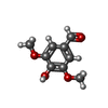  Mass: 182.173 Da / Num. of mol.: 1 / Source method: obtained synthetically / Formula: C9H10O4 / Feature type: SUBJECT OF INVESTIGATION Mass: 182.173 Da / Num. of mol.: 1 / Source method: obtained synthetically / Formula: C9H10O4 / Feature type: SUBJECT OF INVESTIGATION#5: Water | ChemComp-HOH / |  Mass: 18.015 Da / Num. of mol.: 693 / Source method: isolated from a natural source / Formula: H2O Mass: 18.015 Da / Num. of mol.: 693 / Source method: isolated from a natural source / Formula: H2OHas ligand of interest | Y | Has protein modification | Y | |
|---|
-Experimental details
-Experiment
| Experiment | Method: X-RAY DIFFRACTION / Number of used crystals: 1 |
|---|
- Sample preparation
| Crystal | Density Matthews: 2.28 Å3/Da / Density % sol: 46.06 % |
|---|---|
| Crystal grow | Temperature: 291 K / Method: vapor diffusion, hanging drop / pH: 6.5 Details: 12-15.5% PEG 3350, 0.1M MgCl2, 0.1M pH 6.5 Bis-tris, VAPOR DIFFUSION, HANGING DROP, temperature 291K |
-Data collection
| Diffraction | Mean temperature: 100 K / Serial crystal experiment: N |
|---|---|
| Diffraction source | Source: SYNCHROTRON / Site: NFPSS  / Beamline: BL19U1 / Wavelength: 0.979 Å / Beamline: BL19U1 / Wavelength: 0.979 Å |
| Detector | Type: DECTRIS PILATUS 6M / Detector: PIXEL / Date: Jan 1, 2022 |
| Radiation | Protocol: SINGLE WAVELENGTH / Monochromatic (M) / Laue (L): M / Scattering type: x-ray |
| Radiation wavelength | Wavelength: 0.979 Å / Relative weight: 1 |
| Reflection | Resolution: 2→48.6 Å / Num. obs: 70329 / % possible obs: 99.4 % / Redundancy: 4.4 % / CC1/2: 0.983 / Rmerge(I) obs: 0.062 / Net I/σ(I): 12.9 |
| Reflection shell | Resolution: 2→2.04 Å / Rmerge(I) obs: 0.426 / Mean I/σ(I) obs: 3.3 / Num. unique obs: 5420 / CC1/2: 0.875 |
- Processing
| Software |
| ||||||||||||||||||||||||||||||||||||||||||||||||||||||||||||||||||||||||||||||||||||||||||||||||||||||||||||||||||||||||||||||||||||||||||||||||||||||||||||||||||||||||||||||||||||||
|---|---|---|---|---|---|---|---|---|---|---|---|---|---|---|---|---|---|---|---|---|---|---|---|---|---|---|---|---|---|---|---|---|---|---|---|---|---|---|---|---|---|---|---|---|---|---|---|---|---|---|---|---|---|---|---|---|---|---|---|---|---|---|---|---|---|---|---|---|---|---|---|---|---|---|---|---|---|---|---|---|---|---|---|---|---|---|---|---|---|---|---|---|---|---|---|---|---|---|---|---|---|---|---|---|---|---|---|---|---|---|---|---|---|---|---|---|---|---|---|---|---|---|---|---|---|---|---|---|---|---|---|---|---|---|---|---|---|---|---|---|---|---|---|---|---|---|---|---|---|---|---|---|---|---|---|---|---|---|---|---|---|---|---|---|---|---|---|---|---|---|---|---|---|---|---|---|---|---|---|---|---|---|---|
| Refinement | Method to determine structure: MOLECULAR REPLACEMENT Starting model: 4Q89 Resolution: 2→43.85 Å / SU ML: 0.22 / Cross valid method: THROUGHOUT / σ(F): 1.34 / Phase error: 22.67 / Stereochemistry target values: ML
| ||||||||||||||||||||||||||||||||||||||||||||||||||||||||||||||||||||||||||||||||||||||||||||||||||||||||||||||||||||||||||||||||||||||||||||||||||||||||||||||||||||||||||||||||||||||
| Solvent computation | Shrinkage radii: 0.9 Å / VDW probe radii: 1.1 Å / Solvent model: FLAT BULK SOLVENT MODEL | ||||||||||||||||||||||||||||||||||||||||||||||||||||||||||||||||||||||||||||||||||||||||||||||||||||||||||||||||||||||||||||||||||||||||||||||||||||||||||||||||||||||||||||||||||||||
| Displacement parameters | Biso max: 91.63 Å2 / Biso mean: 32.5442 Å2 / Biso min: 12.92 Å2 | ||||||||||||||||||||||||||||||||||||||||||||||||||||||||||||||||||||||||||||||||||||||||||||||||||||||||||||||||||||||||||||||||||||||||||||||||||||||||||||||||||||||||||||||||||||||
| Refinement step | Cycle: final / Resolution: 2→43.85 Å
| ||||||||||||||||||||||||||||||||||||||||||||||||||||||||||||||||||||||||||||||||||||||||||||||||||||||||||||||||||||||||||||||||||||||||||||||||||||||||||||||||||||||||||||||||||||||
| LS refinement shell | Refine-ID: X-RAY DIFFRACTION / Rfactor Rfree error: 0 / Total num. of bins used: 25
|
