 Movie
Movie Controller

[English] 日本語
 Yorodumi
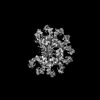
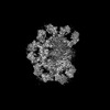
- PDB-7y0h: Cryo-EM structure of human IgM-Fc in complex with the J chain and... -
+ Open data

- Basic information
| Entry | Database: PDB / ID: 7y0h | |||||||||||||||||||||||||||||||||||||||||||||||||||||||||
|---|---|---|---|---|---|---|---|---|---|---|---|---|---|---|---|---|---|---|---|---|---|---|---|---|---|---|---|---|---|---|---|---|---|---|---|---|---|---|---|---|---|---|---|---|---|---|---|---|---|---|---|---|---|---|---|---|---|---|
| Title | Cryo-EM structure of human IgM-Fc in complex with the J chain and the P. falciparum VAR2CSA FCR3 | |||||||||||||||||||||||||||||||||||||||||||||||||||||||||
 Components Components |
| |||||||||||||||||||||||||||||||||||||||||||||||||||||||||
 Keywords Keywords | IMMUNE SYSTEM / Complex / antibody | |||||||||||||||||||||||||||||||||||||||||||||||||||||||||
| Function / homology |  Function and homology information Function and homology informationhexameric IgM immunoglobulin complex / dimeric IgA immunoglobulin complex / IgM B cell receptor complex / secretory dimeric IgA immunoglobulin complex / monomeric IgA immunoglobulin complex / pentameric IgM immunoglobulin complex / secretory IgA immunoglobulin complex / IgA binding / IgM immunoglobulin complex / glomerular filtration ...hexameric IgM immunoglobulin complex / dimeric IgA immunoglobulin complex / IgM B cell receptor complex / secretory dimeric IgA immunoglobulin complex / monomeric IgA immunoglobulin complex / pentameric IgM immunoglobulin complex / secretory IgA immunoglobulin complex / IgA binding / IgM immunoglobulin complex / glomerular filtration / pre-B cell allelic exclusion / CD22 mediated BCR regulation / immunoglobulin receptor binding / positive regulation of respiratory burst / humoral immune response / Scavenging of heme from plasma / antigen binding / Antigen activates B Cell Receptor (BCR) leading to generation of second messengers / Cell surface interactions at the vascular wall / B cell receptor signaling pathway / antibacterial humoral response / protein-containing complex assembly / blood microparticle / Potential therapeutics for SARS / defense response to Gram-negative bacterium / adaptive immune response / protein-macromolecule adaptor activity / host cell surface receptor binding / immune response / innate immune response / cell surface / protein homodimerization activity / : / extracellular exosome / extracellular region / membrane / plasma membrane Similarity search - Function | |||||||||||||||||||||||||||||||||||||||||||||||||||||||||
| Biological species |  Homo sapiens (human) Homo sapiens (human) | |||||||||||||||||||||||||||||||||||||||||||||||||||||||||
| Method | ELECTRON MICROSCOPY / single particle reconstruction / cryo EM / Resolution: 3.56 Å | |||||||||||||||||||||||||||||||||||||||||||||||||||||||||
 Authors Authors | Ji, C. / Xiao, J. | |||||||||||||||||||||||||||||||||||||||||||||||||||||||||
| Funding support |  China, 1items China, 1items
| |||||||||||||||||||||||||||||||||||||||||||||||||||||||||
 Citation Citation | Journal: Nat Commun / Year: 2023 Title: Plasmodium falciparum has evolved multiple mechanisms to hijack human immunoglobulin M. Authors: Chenggong Ji / Hao Shen / Chen Su / Yaxin Li / Shihua Chen / Thomas H Sharp / Junyu Xiao /  Abstract: Plasmodium falciparum causes the most severe malaria in humans. Immunoglobulin M (IgM) serves as the first line of humoral defense against infection and potently activates the complement pathway to ...Plasmodium falciparum causes the most severe malaria in humans. Immunoglobulin M (IgM) serves as the first line of humoral defense against infection and potently activates the complement pathway to facilitate P. falciparum clearance. A number of P. falciparum proteins bind IgM, leading to immune evasion and severe disease. However, the underlying molecular mechanisms remain unknown. Here, using high-resolution cryo-electron microscopy, we delineate how P. falciparum proteins VAR2CSA, TM284VAR1, DBLMSP, and DBLMSP2 target IgM. Each protein binds IgM in a different manner, and together they present a variety of Duffy-binding-like domain-IgM interaction modes. We further show that these proteins interfere directly with IgM-mediated complement activation in vitro, with VAR2CSA exhibiting the most potent inhibitory effect. These results underscore the importance of IgM for human adaptation of P. falciparum and provide critical insights into its immune evasion mechanism. | |||||||||||||||||||||||||||||||||||||||||||||||||||||||||
| History |
|
- Structure visualization
| Structure viewer | Molecule: MolmilJmol/JSmol |
|---|
- Downloads & links
-Download
| PDBx/mmCIF format | 7y0h.cif.gz | 641.9 KB | Display | PDBx/mmCIF format |
|---|---|---|---|---|
| PDB format | pdb7y0h.ent.gz | 497 KB | Display | PDB format |
| PDBx/mmJSON format | 7y0h.json.gz | Tree view | PDBx/mmJSON format | |
| Others |  Other downloads Other downloads |
-Validation report
| Arichive directory | https://data.pdbj.org/pub/pdb/validation_reports/y0/7y0hftp://data.pdbj.org/pub/pdb/validation_reports/y0/7y0h | HTTPS FTP |
|---|
-Related structure data
| Related structure data | 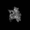 33542MC  7y09C  7y0jC  7yg2C M: map data used to model this data C: citing same article ( |
|---|---|
| Similar structure data |


-Links
 PDBj
PDBj


- Assembly
| Deposited unit | 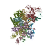
|
|---|---|
| 1 |
|
-Components
| #1: Protein | Mass: 41875.766 Da / Num. of mol.: 10 Source method: isolated from a genetically manipulated source Source: (gene. exp.) Homo sapiens (human) / Gene: IGHM / Cell line (production host): HEK293 / Production host: Homo sapiens (human) / References: UniProt: P01871#2: Protein | | Mass: 15483.329 Da / Num. of mol.: 1 Source method: isolated from a genetically manipulated source Source: (gene. exp.) Homo sapiens (human) / Gene: JCHAIN, IGCJ, IGJ / Cell line (production host): HEK293 / Production host: Homo sapiens (human) / References: UniProt: P01591#3: Protein | | Mass: 306371.406 Da / Num. of mol.: 1 Source method: isolated from a genetically manipulated source Source: (gene. exp.) Gene: var / Production host:  Trichoplusia ni (cabbage looper) / References: UniProt: Q6UDW7 Trichoplusia ni (cabbage looper) / References: UniProt: Q6UDW7#4: Polysaccharide | 2-acetamido-2-deoxy-beta-D-glucopyranose-(1-4)-2-acetamido-2-deoxy-beta-D-glucopyranose | Source method: isolated from a genetically manipulated source #5: Sugar | ChemComp-NAG /   Type: D-saccharide, beta linking / Mass: 221.208 Da / Num. of mol.: 10 / Source method: obtained synthetically / Formula: C8H15NO6 / Feature type: SUBJECT OF INVESTIGATION Type: D-saccharide, beta linking / Mass: 221.208 Da / Num. of mol.: 10 / Source method: obtained synthetically / Formula: C8H15NO6 / Feature type: SUBJECT OF INVESTIGATIONHas ligand of interest | Y | Has protein modification | Y | |
|---|
-Experimental details
-Experiment
| Experiment | Method: ELECTRON MICROSCOPY |
|---|---|
| EM experiment | Aggregation state: PARTICLE / 3D reconstruction method: single particle reconstruction |
- Sample preparation
| Component |
| ||||||||||||||||||||||||
|---|---|---|---|---|---|---|---|---|---|---|---|---|---|---|---|---|---|---|---|---|---|---|---|---|---|
| Source (natural) |
| ||||||||||||||||||||||||
| Source (recombinant) |
| ||||||||||||||||||||||||
| Buffer solution | pH: 7.4 | ||||||||||||||||||||||||
| Specimen | Embedding applied: NO / Shadowing applied: NO / Staining applied: NO / Vitrification applied: YES | ||||||||||||||||||||||||
| Vitrification | Cryogen name: ETHANE |
- Electron microscopy imaging
| Experimental equipment |  Model: Titan Krios / Image courtesy: FEI Company |
|---|---|
| Microscopy | Model: FEI TITAN KRIOS |
| Electron gun | Electron source:  FIELD EMISSION GUN / Accelerating voltage: 300 kV / Illumination mode: FLOOD BEAM FIELD EMISSION GUN / Accelerating voltage: 300 kV / Illumination mode: FLOOD BEAM |
| Electron lens | Mode: BRIGHT FIELD / Nominal defocus max: 2000 nm / Nominal defocus min: 1000 nm |
| Image recording | Electron dose: 60 e/Å2 / Film or detector model: GATAN K3 (6k x 4k) |
- Processing
| Software | Name: PHENIX / Version: 1.19.1_4122: / Classification: refinement | ||||||||||||||||||||||||
|---|---|---|---|---|---|---|---|---|---|---|---|---|---|---|---|---|---|---|---|---|---|---|---|---|---|
| EM software | Name: PHENIX / Category: model refinement | ||||||||||||||||||||||||
| CTF correction | Type: PHASE FLIPPING AND AMPLITUDE CORRECTION | ||||||||||||||||||||||||
| Symmetry | Point symmetry: C1 (asymmetric) | ||||||||||||||||||||||||
| 3D reconstruction | Resolution: 3.56 Å / Resolution method: FSC 0.143 CUT-OFF / Num. of particles: 690345 / Symmetry type: POINT | ||||||||||||||||||||||||
| Refine LS restraints |
|
