 Movie
Movie Controller


+ Open data

- Basic information
| Entry | Database: PDB / ID: 7xzf | ||||||||||||
|---|---|---|---|---|---|---|---|---|---|---|---|---|---|
| Title | Wild type of the N-terminal domain of fucoidan lyase FdlA | ||||||||||||
 Components Components | Fucoidan lyase | ||||||||||||
 Keywords Keywords | LYASE / Fucobacter marina / fucoidan lyase / polysaccharide lyase | ||||||||||||
| Function / homology | IMIDAZOLE Function and homology information Function and homology information | ||||||||||||
| Biological species |  Flavobacteriaceae bacterium SA-0082 (bacteria) Flavobacteriaceae bacterium SA-0082 (bacteria) | ||||||||||||
| Method |  X-RAY DIFFRACTION / SYNCHROTRON / SAD / Resolution: 1.3 Å X-RAY DIFFRACTION / SYNCHROTRON / SAD / Resolution: 1.3 Å | ||||||||||||
 Authors Authors | Wang, J. / Li, M. / Pan, X. | ||||||||||||
| Funding support |  China, 3items China, 3items
| ||||||||||||
 Citation Citation | Journal: Mar Drugs / Year: 2022 Title: Structural and Biochemical Analysis Reveals Catalytic Mechanism of Fucoidan Lyase from Flavobacterium sp. SA-0082. Authors: Wang, J. / Liu, Z. / Pan, X. / Wang, N. / Li, L. / Du, Y. / Li, J. / Li, M. | ||||||||||||
| History |
|
- Structure visualization
| Structure viewer | Molecule: MolmilJmol/JSmol |
|---|
- Downloads & links
-Download
| PDBx/mmCIF format | 7xzf.cif.gz | 450.3 KB | Display | PDBx/mmCIF format |
|---|---|---|---|---|
| PDB format | pdb7xzf.ent.gz | 327 KB | Display | PDB format |
| PDBx/mmJSON format | 7xzf.json.gz | Tree view | PDBx/mmJSON format | |
| Others |  Other downloads Other downloads |
-Validation report
| Arichive directory | https://data.pdbj.org/pub/pdb/validation_reports/xz/7xzfftp://data.pdbj.org/pub/pdb/validation_reports/xz/7xzf | HTTPS FTP |
|---|
-Related structure data
| Related structure data |  7xz7C  7xz8C  7xz9C 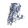 7xzaC  7xzbC 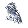 7xzcC  7xzdC  7xzeC C: citing same article ( |
|---|---|
| Similar structure data |
-Links
 PDBj
PDBj


- Assembly
| Deposited unit | 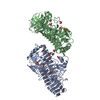
| ||||||||||||
|---|---|---|---|---|---|---|---|---|---|---|---|---|---|
| 1 | 
| ||||||||||||
| 2 | 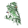
| ||||||||||||
| Unit cell |
|
-Components
| #1: Protein | Mass: 50256.434 Da / Num. of mol.: 2 / Fragment: N-terminal domain Source method: isolated from a genetically manipulated source Details: Genbank AAO00510.1,Sequence 3 from patent US 6489155 Source: (gene. exp.) Flavobacteriaceae bacterium SA-0082 (bacteria)Gene: AAO5510.1 / Production host: References: Lyases; Carbon-oxygen lyases; Acting on polysaccharides #2: Chemical | ChemComp-SO4 /   Mass: 96.063 Da / Num. of mol.: 9 / Source method: obtained synthetically / Formula: SO4 Mass: 96.063 Da / Num. of mol.: 9 / Source method: obtained synthetically / Formula: SO4#3: Chemical |   Mass: 69.085 Da / Num. of mol.: 2 / Source method: obtained synthetically / Formula: C3H5N2 Mass: 69.085 Da / Num. of mol.: 2 / Source method: obtained synthetically / Formula: C3H5N2#4: Chemical | ChemComp-GOL / |   Mass: 92.094 Da / Num. of mol.: 1 / Source method: obtained synthetically / Formula: C3H8O3 Mass: 92.094 Da / Num. of mol.: 1 / Source method: obtained synthetically / Formula: C3H8O3#5: Water | ChemComp-HOH / |  Mass: 18.015 Da / Num. of mol.: 962 / Source method: isolated from a natural source / Formula: H2O Mass: 18.015 Da / Num. of mol.: 962 / Source method: isolated from a natural source / Formula: H2OHas ligand of interest | N | |
|---|
-Experimental details
-Experiment
| Experiment | Method: X-RAY DIFFRACTION / Number of used crystals: 1 |
|---|
- Sample preparation
| Crystal | Density Matthews: 2.81 Å3/Da / Density % sol: 56.19 % |
|---|---|
| Crystal grow | Temperature: 291.15 K / Method: vapor diffusion, sitting drop Details: 0.1M CAPS pH 10.5, 0.2M Lithium sulfate, 2M ammonium sulfate PH range: 7.5-10.5 |
-Data collection
| Diffraction | Mean temperature: 193.15 K / Serial crystal experiment: N |
|---|---|
| Diffraction source | Source: SYNCHROTRON / Site: SSRF / Beamline: BL17U1 / Wavelength: 0.9785 Å |
| Detector | Type: DECTRIS EIGER X 16M / Detector: PIXEL / Date: Dec 29, 2017 |
| Radiation | Protocol: SINGLE WAVELENGTH / Monochromatic (M) / Laue (L): M / Scattering type: x-ray |
| Radiation wavelength | Wavelength: 0.9785 Å / Relative weight: 1 |
| Reflection | Resolution: 1.25→50 Å / Num. obs: 290039 / % possible obs: 94.8 % / Redundancy: 2 % / Biso Wilson estimate: 8.82 Å2 / Rrim(I) all: 0.113 / Net I/σ(I): 8.9 |
| Reflection shell | Resolution: 1.25→1.27 Å / Num. unique obs: 13327 / Rpim(I) all: 0.505 |
- Processing
| Software |
| |||||||||||||||||||||||||||||||||||||||||||||||||||||||||||||||||||||||||||||||||||||||||||||||||||||||||||||||||||||||||||||||||||||||||||||||||||||||||||||||||||||||||||||||||||||||||||||||||||||||||||||||||||||||||
|---|---|---|---|---|---|---|---|---|---|---|---|---|---|---|---|---|---|---|---|---|---|---|---|---|---|---|---|---|---|---|---|---|---|---|---|---|---|---|---|---|---|---|---|---|---|---|---|---|---|---|---|---|---|---|---|---|---|---|---|---|---|---|---|---|---|---|---|---|---|---|---|---|---|---|---|---|---|---|---|---|---|---|---|---|---|---|---|---|---|---|---|---|---|---|---|---|---|---|---|---|---|---|---|---|---|---|---|---|---|---|---|---|---|---|---|---|---|---|---|---|---|---|---|---|---|---|---|---|---|---|---|---|---|---|---|---|---|---|---|---|---|---|---|---|---|---|---|---|---|---|---|---|---|---|---|---|---|---|---|---|---|---|---|---|---|---|---|---|---|---|---|---|---|---|---|---|---|---|---|---|---|---|---|---|---|---|---|---|---|---|---|---|---|---|---|---|---|---|---|---|---|---|---|---|---|---|---|---|---|---|---|---|---|---|---|---|---|---|
| Refinement | Method to determine structure: SAD / Resolution: 1.3→24.99 Å / SU ML: 0.1097 / Cross valid method: FREE R-VALUE / σ(F): 1.97 / Phase error: 14.9068 Stereochemistry target values: GeoStd + Monomer Library + CDL v1.2
| |||||||||||||||||||||||||||||||||||||||||||||||||||||||||||||||||||||||||||||||||||||||||||||||||||||||||||||||||||||||||||||||||||||||||||||||||||||||||||||||||||||||||||||||||||||||||||||||||||||||||||||||||||||||||
| Solvent computation | Shrinkage radii: 0.9 Å / VDW probe radii: 1.11 Å / Solvent model: FLAT BULK SOLVENT MODEL | |||||||||||||||||||||||||||||||||||||||||||||||||||||||||||||||||||||||||||||||||||||||||||||||||||||||||||||||||||||||||||||||||||||||||||||||||||||||||||||||||||||||||||||||||||||||||||||||||||||||||||||||||||||||||
| Displacement parameters | Biso mean: 12.94 Å2 | |||||||||||||||||||||||||||||||||||||||||||||||||||||||||||||||||||||||||||||||||||||||||||||||||||||||||||||||||||||||||||||||||||||||||||||||||||||||||||||||||||||||||||||||||||||||||||||||||||||||||||||||||||||||||
| Refinement step | Cycle: LAST / Resolution: 1.3→24.99 Å
| |||||||||||||||||||||||||||||||||||||||||||||||||||||||||||||||||||||||||||||||||||||||||||||||||||||||||||||||||||||||||||||||||||||||||||||||||||||||||||||||||||||||||||||||||||||||||||||||||||||||||||||||||||||||||
| Refine LS restraints |
| |||||||||||||||||||||||||||||||||||||||||||||||||||||||||||||||||||||||||||||||||||||||||||||||||||||||||||||||||||||||||||||||||||||||||||||||||||||||||||||||||||||||||||||||||||||||||||||||||||||||||||||||||||||||||
| LS refinement shell |
|
