 Movie
Movie Controller

[English] 日本語
 Yorodumi
- PDB-7xy8: Crystal structure of antibody Fab fragment in complex with CD147(... -
+ Open data

- Basic information
| Entry | Database: PDB / ID: 7xy8 | ||||||
|---|---|---|---|---|---|---|---|
| Title | Crystal structure of antibody Fab fragment in complex with CD147(EMMPIRIN) | ||||||
 Components Components |
| ||||||
 Keywords Keywords | PROTEIN BINDING / cd147 / complex / antibody | ||||||
| Function / homology |  Function and homology information Function and homology informationDefective SLC16A1 causes symptomatic deficiency in lactate transport (SDLT) / Proton-coupled monocarboxylate transport / positive regulation of matrix metallopeptidase secretion / dendrite self-avoidance / acrosomal membrane / cell-cell adhesion mediator activity / endothelial tube morphogenesis / response to mercury ion / neural retina development / photoreceptor cell maintenance ...Defective SLC16A1 causes symptomatic deficiency in lactate transport (SDLT) / Proton-coupled monocarboxylate transport / positive regulation of matrix metallopeptidase secretion / dendrite self-avoidance / acrosomal membrane / cell-cell adhesion mediator activity / endothelial tube morphogenesis / response to mercury ion / neural retina development / photoreceptor cell maintenance / Basigin interactions / Aspirin ADME / D-mannose binding / homophilic cell-cell adhesion / odontogenesis of dentin-containing tooth / decidualization / positive regulation of vascular endothelial growth factor production / photoreceptor outer segment / response to cAMP / Integrin cell surface interactions / Degradation of the extracellular matrix / neutrophil chemotaxis / photoreceptor inner segment / embryo implantation / positive regulation of endothelial cell migration / axon guidance / protein localization to plasma membrane / response to peptide hormone / sarcolemma / positive regulation of interleukin-6 production / melanosome / signaling receptor activity / virus receptor activity / angiogenesis / basolateral plasma membrane / positive regulation of viral entry into host cell / cell surface receptor signaling pathway / endosome / cadherin binding / Golgi membrane / axon / focal adhesion / endoplasmic reticulum membrane / mitochondrion / extracellular exosome / membrane / plasma membrane Similarity search - Function | ||||||
| Biological species |  Homo sapiens (human) Homo sapiens (human)synthetic construct (others) | ||||||
| Method |  X-RAY DIFFRACTION / SYNCHROTRON / MOLECULAR REPLACEMENT / Resolution: 2.3 Å X-RAY DIFFRACTION / SYNCHROTRON / MOLECULAR REPLACEMENT / Resolution: 2.3 Å | ||||||
 Authors Authors | Nakamura, K. / Amano, M. / Yoneda, K. / Suzuki, M. / Fukuchi, K. | ||||||
| Funding support | 1items
| ||||||
 Citation Citation | Journal: J Oncol / Year: 2022 Title: Novel Antibody Exerts Antitumor Effect through Downregulation of CD147 and Activation of Multiple Stress Signals. Authors: Fukuchi, K. / Nanai, K. / Yuita, H. / Maru, C. / Tsukada, J. / Ishigami, M. / Nagai, Y. / Nakano, Y. / Yoshimura, C. / Yoneda, K. / Amano, M. / Nakamura, K. / Oda, Y. / Nishigohri, H. / ...Authors: Fukuchi, K. / Nanai, K. / Yuita, H. / Maru, C. / Tsukada, J. / Ishigami, M. / Nagai, Y. / Nakano, Y. / Yoshimura, C. / Yoneda, K. / Amano, M. / Nakamura, K. / Oda, Y. / Nishigohri, H. / Yamamoto, S. / Ohnishi-Totoki, Y. / Inaki, K. / Komori, H. / Nakano, R. / Kanari, Y. / Nishida, A. / Matsui, Y. / Funo, S. / Takahashi, S. / Ohtsuka, T. / Agatsuma, T. | ||||||
| History |
|
- Structure visualization
| Structure viewer | Molecule: MolmilJmol/JSmol |
|---|
- Downloads & links
-Download
| PDBx/mmCIF format | 7xy8.cif.gz | 232.3 KB | Display | PDBx/mmCIF format |
|---|---|---|---|---|
| PDB format | pdb7xy8.ent.gz | 183.7 KB | Display | PDB format |
| PDBx/mmJSON format | 7xy8.json.gz | Tree view | PDBx/mmJSON format | |
| Others |  Other downloads Other downloads |
-Validation report
| Arichive directory | https://data.pdbj.org/pub/pdb/validation_reports/xy/7xy8ftp://data.pdbj.org/pub/pdb/validation_reports/xy/7xy8 | HTTPS FTP |
|---|
-Related structure data
| Related structure data | 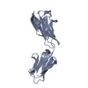 3b5hS S: Starting model for refinement |
|---|---|
| Similar structure data |
-Links
 PDBj
PDBj


- Assembly
| Deposited unit | 
| |||||||||||||||||||||||||||||||||||||||||||||||||||||||||||||||||||||||||||||||||||||||||||||||
|---|---|---|---|---|---|---|---|---|---|---|---|---|---|---|---|---|---|---|---|---|---|---|---|---|---|---|---|---|---|---|---|---|---|---|---|---|---|---|---|---|---|---|---|---|---|---|---|---|---|---|---|---|---|---|---|---|---|---|---|---|---|---|---|---|---|---|---|---|---|---|---|---|---|---|---|---|---|---|---|---|---|---|---|---|---|---|---|---|---|---|---|---|---|---|---|---|
| 1 | 
| |||||||||||||||||||||||||||||||||||||||||||||||||||||||||||||||||||||||||||||||||||||||||||||||
| 2 | 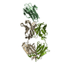
| |||||||||||||||||||||||||||||||||||||||||||||||||||||||||||||||||||||||||||||||||||||||||||||||
| Unit cell |
| |||||||||||||||||||||||||||||||||||||||||||||||||||||||||||||||||||||||||||||||||||||||||||||||
| Noncrystallographic symmetry (NCS) | NCS domain:
NCS domain segments: Component-ID: _ / Refine code: _
NCS ensembles :
|
-Components
| #1: Protein | Mass: 22503.928 Da / Num. of mol.: 2 Source method: isolated from a genetically manipulated source Source: (gene. exp.) Homo sapiens (human) / Gene: BSG, UNQ6505/PRO21383 / Production host:  #2: Antibody | Mass: 25277.320 Da / Num. of mol.: 2 Source method: isolated from a genetically manipulated source Source: (gene. exp.) synthetic construct (others) / Production host: Homo sapiens (human)#3: Antibody | Mass: 23432.900 Da / Num. of mol.: 2 Source method: isolated from a genetically manipulated source Source: (gene. exp.) synthetic construct (others) / Production host: Homo sapiens (human)#4: Water | ChemComp-HOH / |  Mass: 18.015 Da / Num. of mol.: 187 / Source method: isolated from a natural source / Formula: H2O Mass: 18.015 Da / Num. of mol.: 187 / Source method: isolated from a natural source / Formula: H2OHas protein modification | Y | |
|---|
-Experimental details
-Experiment
| Experiment | Method: X-RAY DIFFRACTION / Number of used crystals: 1 |
|---|
- Sample preparation
| Crystal | Density Matthews: 2.09 Å3/Da / Density % sol: 41.23 % |
|---|---|
| Crystal grow | Temperature: 293 K / Method: vapor diffusion / pH: 7 Details: 0.1M Sodium Malonate pH 7.0, 12% (w/v) Polyethylene Glycol 3350 |
-Data collection
| Diffraction | Mean temperature: 100 K / Ambient temp details: Nitrogen gas flow (95K-300K) / Serial crystal experiment: N |
|---|---|
| Diffraction source | Source: SYNCHROTRON / Site: Photon Factory  / Beamline: BL-17A / Wavelength: 0.98 Å / Beamline: BL-17A / Wavelength: 0.98 Å |
| Detector | Type: DECTRIS PILATUS 6M / Detector: PIXEL / Date: Jan 25, 2018 |
| Radiation | Monochromator: Si(111) double crystal / Protocol: SINGLE WAVELENGTH / Monochromatic (M) / Laue (L): M / Scattering type: x-ray |
| Radiation wavelength | Wavelength: 0.98 Å / Relative weight: 1 |
| Reflection | Resolution: 2.3→35.911 Å / Num. obs: 51301 / % possible obs: 98.4 % / Redundancy: 3 % / Rpim(I) all: 0.06 / Rrim(I) all: 0.105 / Rsym value: 0.086 / Net I/av σ(I): 5.4 / Net I/σ(I): 7.8 |
| Reflection shell | Resolution: 2.3→2.42 Å / Redundancy: 3 % / Rmerge(I) obs: 0.36 / Mean I/σ(I) obs: 2.1 / Num. unique obs: 7430 / Rpim(I) all: 0.247 / Rrim(I) all: 0.439 / Rsym value: 0.36 / % possible all: 97.8 |
- Processing
| Software |
| |||||||||||||||||||||||||||||||||||||||||||||
|---|---|---|---|---|---|---|---|---|---|---|---|---|---|---|---|---|---|---|---|---|---|---|---|---|---|---|---|---|---|---|---|---|---|---|---|---|---|---|---|---|---|---|---|---|---|---|
| Refinement | Method to determine structure: MOLECULAR REPLACEMENT Starting model: 3b5h Resolution: 2.3→35.91 Å / Cor.coef. Fo:Fc: 0.92 / Cor.coef. Fo:Fc free: 0.879 / Cross valid method: THROUGHOUT / σ(F): 0 / ESU R: 0.455 / ESU R Free: 0.282 / Stereochemistry target values: MAXIMUM LIKELIHOOD / Details: U VALUES : REFINED INDIVIDUALLY
| |||||||||||||||||||||||||||||||||||||||||||||
| Solvent computation | Ion probe radii: 0.8 Å / Shrinkage radii: 0.8 Å / VDW probe radii: 1.2 Å / Solvent model: MASK | |||||||||||||||||||||||||||||||||||||||||||||
| Displacement parameters | Biso max: 143.51 Å2 / Biso mean: 43.373 Å2 / Biso min: 15.57 Å2
| |||||||||||||||||||||||||||||||||||||||||||||
| Refinement step | Cycle: final / Resolution: 2.3→35.91 Å
| |||||||||||||||||||||||||||||||||||||||||||||
| Refine LS restraints |
| |||||||||||||||||||||||||||||||||||||||||||||
| Refine LS restraints NCS | Refine-ID: X-RAY DIFFRACTION / Type: interatomic distance / Weight position: 0.05
| |||||||||||||||||||||||||||||||||||||||||||||
| LS refinement shell | Resolution: 2.3→2.359 Å / Rfactor Rfree error: 0 / Total num. of bins used: 20
|
