Movie
Movie Controller

[English] 日本語
 Yorodumi
- PDB-7xxf: Structure of photosynthetic LH1-RC super-complex of Rhodopila glo... -
+ Open data

- Basic information
| Entry | Database: PDB / ID: 7xxf | |||||||||||||||||||||||||||||||||||||||
|---|---|---|---|---|---|---|---|---|---|---|---|---|---|---|---|---|---|---|---|---|---|---|---|---|---|---|---|---|---|---|---|---|---|---|---|---|---|---|---|---|
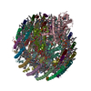
| Title | Structure of photosynthetic LH1-RC super-complex of Rhodopila globiformis | |||||||||||||||||||||||||||||||||||||||
 Components Components |
| |||||||||||||||||||||||||||||||||||||||
 Keywords Keywords | PHOTOSYNTHESIS / LH1-RC COMPLEX / PURPLE BACTERIA | |||||||||||||||||||||||||||||||||||||||
| Function / homology |  Function and homology information Function and homology informationorganelle inner membrane / plasma membrane-derived chromatophore membrane / plasma membrane light-harvesting complex / bacteriochlorophyll binding / photosynthetic electron transport in photosystem II / photosynthesis, light reaction / endomembrane system / electron transfer activity / iron ion binding / heme binding ...organelle inner membrane / plasma membrane-derived chromatophore membrane / plasma membrane light-harvesting complex / bacteriochlorophyll binding / photosynthetic electron transport in photosystem II / photosynthesis, light reaction / endomembrane system / electron transfer activity / iron ion binding / heme binding / metal ion binding / plasma membrane Similarity search - Function | |||||||||||||||||||||||||||||||||||||||
| Biological species |  Rhodopila globiformis (bacteria) Rhodopila globiformis (bacteria) | |||||||||||||||||||||||||||||||||||||||
| Method | ELECTRON MICROSCOPY / single particle reconstruction / cryo EM / Resolution: 2.24 Å | |||||||||||||||||||||||||||||||||||||||
 Authors Authors | Tani, K. / Kanno, R. / Kurosawa, K. / Takaichi, S. / Nagashima, K.V.P. / Hall, M. / Yu, L.-J. / Kimura, Y. / Madigan, M.T. / Mizoguchi, A. ...Tani, K. / Kanno, R. / Kurosawa, K. / Takaichi, S. / Nagashima, K.V.P. / Hall, M. / Yu, L.-J. / Kimura, Y. / Madigan, M.T. / Mizoguchi, A. / Humbel, B.M. / Wang-Otomo, Z.-Y. | |||||||||||||||||||||||||||||||||||||||
| Funding support |  Japan, 6items Japan, 6items
| |||||||||||||||||||||||||||||||||||||||
 Citation Citation | Journal: Commun Biol / Year: 2022 Title: An LH1-RC photocomplex from an extremophilic phototroph provides insight into origins of two photosynthesis proteins. Authors: Kazutoshi Tani / Ryo Kanno / Keigo Kurosawa / Shinichi Takaichi / Kenji V P Nagashima / Malgorzata Hall / Long-Jiang Yu / Yukihiro Kimura / Michael T Madigan / Akira Mizoguchi / Bruno M ...Authors: Kazutoshi Tani / Ryo Kanno / Keigo Kurosawa / Shinichi Takaichi / Kenji V P Nagashima / Malgorzata Hall / Long-Jiang Yu / Yukihiro Kimura / Michael T Madigan / Akira Mizoguchi / Bruno M Humbel / Zheng-Yu Wang-Otomo /   Abstract: Rhodopila globiformis is the most acidophilic of anaerobic purple phototrophs, growing optimally in culture at pH 5. Here we present a cryo-EM structure of the light-harvesting 1-reaction center (LH1- ...Rhodopila globiformis is the most acidophilic of anaerobic purple phototrophs, growing optimally in culture at pH 5. Here we present a cryo-EM structure of the light-harvesting 1-reaction center (LH1-RC) complex from Rhodopila globiformis at 2.24 Å resolution. All purple bacterial cytochrome (Cyt, encoded by the gene pufC) subunit-associated RCs with known structures have their N-termini truncated. By contrast, the Rhodopila globiformis RC contains a full-length tetra-heme Cyt with its N-terminus embedded in the membrane forming an α-helix as the membrane anchor. Comparison of the N-terminal regions of the Cyt with PufX polypeptides widely distributed in Rhodobacter species reveals significant structural similarities, supporting a longstanding hypothesis that PufX is phylogenetically related to the N-terminus of the RC-bound Cyt subunit and that a common ancestor of phototrophic Proteobacteria contained a full-length tetra-heme Cyt subunit that evolved independently through partial deletions of its pufC gene. Eleven copies of a novel γ-like polypeptide were also identified in the bacteriochlorophyll a-containing Rhodopila globiformis LH1 complex; γ-polypeptides have previously been found only in the LH1 of bacteriochlorophyll b-containing species. These features are discussed in relation to their predicted functions of stabilizing the LH1 structure and regulating quinone transport under the warm acidic conditions. #1: Journal: Commun Biol / Year: 2022Title: An LH1-RC photocomplex from an extremophilic phototroph provides insight into origins of two photosynthesis proteins Authors: Tani, K. / Kanno, R. / Kurosawa, K. / Takaichi, S. / Nagashima, K.V.P. / Hall, M. / Yu, L.-J. / Kimura, Y. / Madigan, M.T. / Mizoguchi, A. / Humbel, B.M. / Wang-Otomo, Z.-Y. | |||||||||||||||||||||||||||||||||||||||
| History |
|
- Structure visualization
| Structure viewer | Molecule: MolmilJmol/JSmol |
|---|
- Downloads & links
-Download
| PDBx/mmCIF format | 7xxf.cif.gz | 749.9 KB | Display | PDBx/mmCIF format |
|---|---|---|---|---|
| PDB format | pdb7xxf.ent.gz | 631.1 KB | Display | PDB format |
| PDBx/mmJSON format | 7xxf.json.gz | Tree view | PDBx/mmJSON format | |
| Others |  Other downloads Other downloads |
-Validation report
| Arichive directory | https://data.pdbj.org/pub/pdb/validation_reports/xx/7xxfftp://data.pdbj.org/pub/pdb/validation_reports/xx/7xxf | HTTPS FTP |
|---|
-Related structure data
| Related structure data |  33501MC M: map data used to model this data C: citing same article ( |
|---|---|
| Similar structure data |
-Links
 PDBj
PDBj


- Assembly
| Deposited unit | 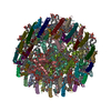
|
|---|---|
| 1 |
|
-Components
-Photosynthetic reaction center ... , 2 types, 2 molecules CH
| #1: Protein | Mass: 37945.188 Da / Num. of mol.: 1 / Source method: isolated from a natural source / Source: (natural) Rhodopila globiformis (bacteria) / References: UniProt: A0A2S6NEK5 |
|---|---|
| #4: Protein | Mass: 28383.367 Da / Num. of mol.: 1 / Source method: isolated from a natural source / Source: (natural) Rhodopila globiformis (bacteria) / References: UniProt: A0A2S6MZS1 |
-Reaction center protein ... , 2 types, 2 molecules LM
| #2: Protein | Mass: 30697.746 Da / Num. of mol.: 1 / Source method: isolated from a natural source / Source: (natural) Rhodopila globiformis (bacteria) / References: UniProt: A0A2S6NEG7 |
|---|---|
| #3: Protein | Mass: 36345.332 Da / Num. of mol.: 1 / Source method: isolated from a natural source / Source: (natural) Rhodopila globiformis (bacteria) / References: UniProt: A0A2S6NEP5 |
-Light-harvesting ... , 3 types, 43 molecules ADFIKOQSUWY13579BEGJNPRTVXZ246...
| #5: Protein | Mass: 7048.523 Da / Num. of mol.: 16 / Source method: isolated from a natural source / Source: (natural) Rhodopila globiformis (bacteria) / References: UniProt: A0A2S6NEK3#6: Protein | Mass: 7877.075 Da / Num. of mol.: 16 / Source method: isolated from a natural source / Source: (natural) Rhodopila globiformis (bacteria) / References: UniProt: A0A2S6NEL9#7: Protein/peptide | Mass: 2542.199 Da / Num. of mol.: 11 / Source method: isolated from a natural source / Source: (natural) Rhodopila globiformis (bacteria) |
|---|
-Sugars , 1 types, 5 molecules 
| #13: Sugar | ChemComp-LMT /  Type: D-saccharide / Mass: 510.615 Da / Num. of mol.: 5 / Source method: obtained synthetically / Formula: C24H46O11 / Comment: detergent*YM Type: D-saccharide / Mass: 510.615 Da / Num. of mol.: 5 / Source method: obtained synthetically / Formula: C24H46O11 / Comment: detergent*YM |
|---|
-Non-polymers , 11 types, 631 molecules 


| #8: Chemical | ChemComp-HEC /  Mass: 618.503 Da / Num. of mol.: 4 / Source method: obtained synthetically / Formula: C34H34FeN4O4 Mass: 618.503 Da / Num. of mol.: 4 / Source method: obtained synthetically / Formula: C34H34FeN4O4#9: Chemical | ChemComp-PGV / (  Mass: 749.007 Da / Num. of mol.: 18 / Source method: obtained synthetically / Formula: C40H77O10P / Comment: phospholipid*YM Mass: 749.007 Da / Num. of mol.: 18 / Source method: obtained synthetically / Formula: C40H77O10P / Comment: phospholipid*YM#10: Chemical | ChemComp-BCL /  Mass: 911.504 Da / Num. of mol.: 36 / Source method: obtained synthetically / Formula: C55H74MgN4O6 / Feature type: SUBJECT OF INVESTIGATION Mass: 911.504 Da / Num. of mol.: 36 / Source method: obtained synthetically / Formula: C55H74MgN4O6 / Feature type: SUBJECT OF INVESTIGATION#11: Chemical |  Mass: 889.215 Da / Num. of mol.: 2 / Source method: obtained synthetically / Formula: C55H76N4O6 Mass: 889.215 Da / Num. of mol.: 2 / Source method: obtained synthetically / Formula: C55H76N4O6#12: Chemical | ChemComp-U10 /  Mass: 863.343 Da / Num. of mol.: 4 / Source method: obtained synthetically / Formula: C59H90O4 Mass: 863.343 Da / Num. of mol.: 4 / Source method: obtained synthetically / Formula: C59H90O4#14: Chemical | ChemComp-FE / |  Mass: 55.845 Da / Num. of mol.: 1 / Source method: obtained synthetically / Formula: Fe Mass: 55.845 Da / Num. of mol.: 1 / Source method: obtained synthetically / Formula: Fe#15: Chemical | ChemComp-MQ9 / |  Mass: 785.233 Da / Num. of mol.: 1 / Source method: obtained synthetically / Formula: C56H80O2 Mass: 785.233 Da / Num. of mol.: 1 / Source method: obtained synthetically / Formula: C56H80O2#16: Chemical | ChemComp-I7D / (  Mass: 612.924 Da / Num. of mol.: 17 / Source method: obtained synthetically / Formula: C42H60O3 / Feature type: SUBJECT OF INVESTIGATION Mass: 612.924 Da / Num. of mol.: 17 / Source method: obtained synthetically / Formula: C42H60O3 / Feature type: SUBJECT OF INVESTIGATION#17: Chemical | ChemComp-CDL /  Mass: 1464.043 Da / Num. of mol.: 9 / Source method: obtained synthetically / Formula: C81H156O17P2 / Comment: phospholipid*YM Mass: 1464.043 Da / Num. of mol.: 9 / Source method: obtained synthetically / Formula: C81H156O17P2 / Comment: phospholipid*YM#18: Chemical | ChemComp-PEE / |  Mass: 744.034 Da / Num. of mol.: 1 / Source method: obtained synthetically / Formula: C41H78NO8P / Comment: DOPE, phospholipid*YM Mass: 744.034 Da / Num. of mol.: 1 / Source method: obtained synthetically / Formula: C41H78NO8P / Comment: DOPE, phospholipid*YM#19: Water | ChemComp-HOH / | Mass: 18.015 Da / Num. of mol.: 538 / Source method: isolated from a natural source / Formula: H2O |
|---|
-Details
| Has ligand of interest | Y |
|---|---|
| Has protein modification | Y |
-Experimental details
-Experiment
| Experiment | Method: ELECTRON MICROSCOPY |
|---|---|
| EM experiment | Aggregation state: PARTICLE / 3D reconstruction method: single particle reconstruction |
- Sample preparation
| Component | Name: Photosynthetic LH1-RC complex from the purple phototrophic bacterium Rhodopila globiformis Type: COMPLEX / Entity ID: #1-#7 / Source: NATURAL |
|---|---|
| Molecular weight | Value: 0.4 MDa / Experimental value: NO |
| Source (natural) | Organism: Rhodopila globiformis (bacteria) |
| Buffer solution | pH: 7.5 |
| Specimen | Conc.: 5 mg/ml / Embedding applied: NO / Shadowing applied: NO / Staining applied: NO / Vitrification applied: YES / Details: This sample was monodisperse. |
| Vitrification | Instrument: LEICA EM GP / Cryogen name: ETHANE / Humidity: 80 % / Chamber temperature: 277 K |
- Electron microscopy imaging
| Experimental equipment |  Model: Titan Krios / Image courtesy: FEI Company |
|---|---|
| Microscopy | Model: FEI TITAN KRIOS |
| Electron gun | Electron source:  FIELD EMISSION GUN / Accelerating voltage: 300 kV / Illumination mode: FLOOD BEAM FIELD EMISSION GUN / Accelerating voltage: 300 kV / Illumination mode: FLOOD BEAM |
| Electron lens | Mode: BRIGHT FIELD / Nominal defocus max: 2800 nm / Nominal defocus min: 600 nm / Alignment procedure: COMA FREE |
| Specimen holder | Cryogen: NITROGEN / Specimen holder model: FEI TITAN KRIOS AUTOGRID HOLDER |
| Image recording | Average exposure time: 38.9 sec. / Electron dose: 40 e/Å2 / Detector mode: COUNTING / Film or detector model: FEI FALCON III (4k x 4k) |
- Processing
| Software | Name: PHENIX / Version: 1.20_4459: / Classification: refinement | ||||||||||||||||||||||||||||||||
|---|---|---|---|---|---|---|---|---|---|---|---|---|---|---|---|---|---|---|---|---|---|---|---|---|---|---|---|---|---|---|---|---|---|
| EM software |
| ||||||||||||||||||||||||||||||||
| CTF correction | Type: PHASE FLIPPING ONLY | ||||||||||||||||||||||||||||||||
| 3D reconstruction | Resolution: 2.24 Å / Resolution method: FSC 0.143 CUT-OFF / Num. of particles: 128119 / Algorithm: FOURIER SPACE / Symmetry type: POINT | ||||||||||||||||||||||||||||||||
| Atomic model building | B value: 40 / Protocol: RIGID BODY FIT / Space: REAL / Target criteria: Correlation coefficient | ||||||||||||||||||||||||||||||||
| Atomic model building | PDB-ID: 5Y5S Accession code: 5Y5S / Source name: PDB / Type: experimental model | ||||||||||||||||||||||||||||||||
| Refine LS restraints |
|