| Entry | Database: PDB / ID: 7xx4
|
|---|
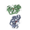
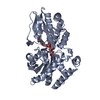
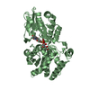
| Title | designed glycosyltransferase |
|---|
 Components Components | Oleandomycin glycosyltransferase |
|---|
 Keywords Keywords | TRANSFERASE / glycosyltransferase |
|---|
| Function / homology |  Function and homology information Function and homology information
UDP-glycosyltransferase, MGT-like / Erythromycin biosynthesis protein CIII-like, central / : / Erythromycin biosynthesis protein CIII-like, C-terminal domain / UDP-glycosyltransferase family, conserved site / UDP-glycosyltransferases signature. / UDP-glucuronosyl/UDP-glucosyltransferaseSimilarity search - Domain/homology |
|---|
| Biological species |  Streptomyces antibioticus (bacteria) Streptomyces antibioticus (bacteria) |
|---|
| Method |  X-RAY DIFFRACTION / SYNCHROTRON / MOLECULAR REPLACEMENT / Resolution: 2.43 Å X-RAY DIFFRACTION / SYNCHROTRON / MOLECULAR REPLACEMENT / Resolution: 2.43 Å |
|---|
 Authors Authors | Lu, M. / Wu, X. |
|---|
| Funding support |  China, 1items China, 1items | Organization | Grant number | Country |
|---|
| National Natural Science Foundation of China (NSFC) | 81973214 | China |
|
|---|
 Citation Citation | Journal: Microb Biotechnol / Year: 2023
Title: Design of a chimeric glycosyltransferase OleD for the site-specific O-monoglycosylation of 3-hydroxypyridine in nosiheptide.
Authors: Zhao, L. / Xu, Y. / Chen, M. / Wu, L. / Li, M. / Lu, Y. / Lu, M. / Chen, Y. / Wu, X. |
|---|
| History | | Deposition | May 28, 2022 | Deposition site: PDBJ / Processing site: PDBJ |
|---|
| Revision 1.0 | Jun 14, 2023 | Provider: repository / Type: Initial release |
|---|
| Revision 1.1 | Nov 29, 2023 | Group: Data collection / Refinement description
Category: chem_comp_atom / chem_comp_bond / pdbx_initial_refinement_model |
|---|
| Revision 1.2 | Dec 27, 2023 | Group: Database references / Category: citation / citation_author
Item: _citation.journal_abbrev / _citation.journal_id_CSD ..._citation.journal_abbrev / _citation.journal_id_CSD / _citation.journal_id_ISSN / _citation.journal_volume / _citation.page_first / _citation.page_last / _citation.pdbx_database_id_DOI / _citation.pdbx_database_id_PubMed / _citation.title / _citation.year |
|---|
|
|---|
 Movie
Movie Controller


 Open data
 Basic information
 Structure visualization
 Downloads & links
 Other downloads
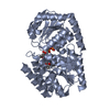
 PDBj
PDBj
 Assembly