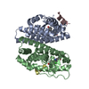
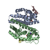
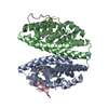
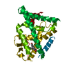
登録情報 データベース : PDB / ID : 7xwqタイトル Human Estrogen Receptor beta Ligand-binding Domain in Complex with (R)-2-(2-chloro-4-hydroxyphenyl)-3-(4-hydroxyphenyl)propanenitrile Estrogen receptor beta SRC peptide キーワード / / / 機能・相同性 分子機能 ドメイン・相同性 構成要素
/ / / / / / / / / / / / / / / / / / / / / / / / / / / / / / / / / / / / / / / / / / / / / / / / / / / / / / / / / / / / / / / / / / / / / 生物種 Homo sapiens (ヒト)手法 / / / 解像度 : 1.89 Å データ登録者 Furuya, N. / Handa, C. 資金援助 1件 ジャーナル : J.Steroid Biochem.Mol.Biol. / 年 : 2022タイトル : Evaluating the correlation of binding affinities between isothermal titration calorimetry and fragment molecular orbital method of estrogen receptor beta with diarylpropionitrile (DPN) or DPN derivatives.著者 : Handa, C. / Yamazaki, Y. / Yonekubo, S. / Furuya, N. / Momose, T. / Ozawa, T. / Furuishi, T. / Fukuzawa, K. / Yonemochi, E. 履歴 登録 2022年5月27日 登録サイト / 処理サイト 改定 1.0 2022年7月20日 Provider / タイプ 改定 1.1 2022年7月27日 Group / Structure summary / カテゴリ / struct / Item / _struct.title改定 1.2 2023年11月29日 Group / Refinement descriptionカテゴリ / chem_comp_bond / pdbx_initial_refinement_model
すべて表示 表示を減らす
 ムービー
ムービー コントローラー


 データを開く
 基本情報
基本情報 要素
要素 キーワード
キーワード 機能・相同性情報
 Homo sapiens (ヒト)
 X線回折 /
  データ登録者
 引用
引用 構造の表示
構造の表示 ダウンロードとリンク
 その他のダウンロード
 PDBj
PDBj
 集合体
集合体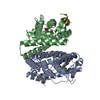

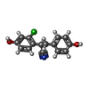
 分子量: 273.714 Da / 分子数: 2 / 由来タイプ: 合成 / 式: C15H12ClNO2
 分子量: 18.015 Da / 分子数: 96 / 由来タイプ: 天然 / 式: H2O
 試料調製
試料調製 / ビームライン: BL-17A / 波長: 1 Å
 解析
解析