 Movie
Movie Controller


+ Open data

- Basic information
| Entry | Database: PDB / ID: 7xw2 | ||||||
|---|---|---|---|---|---|---|---|
| Title | Cryo-EM structure of human DICER-pre-miRNA in a dicing state | ||||||
 Components Components |
| ||||||
 Keywords Keywords | GENE REGULATION/RNA / Dicer / RNaseIII / RNA-binding / micro-RNA processing / GENE REGULATION / GENE REGULATION-RNA complex | ||||||
| Function / homology |  Function and homology information Function and homology informationperipheral nervous system myelin formation / positive regulation of Schwann cell differentiation / global gene silencing by mRNA cleavage / negative regulation of Schwann cell proliferation / tRNA-derived small RNA (tsRNA or tRNA-related fragment, tRF) biogenesis / pre-miRNA binding / tRNA decay / ribonuclease III / Small interfering RNA (siRNA) biogenesis / apoptotic DNA fragmentation ...peripheral nervous system myelin formation / positive regulation of Schwann cell differentiation / global gene silencing by mRNA cleavage / negative regulation of Schwann cell proliferation / tRNA-derived small RNA (tsRNA or tRNA-related fragment, tRF) biogenesis / pre-miRNA binding / tRNA decay / ribonuclease III / Small interfering RNA (siRNA) biogenesis / apoptotic DNA fragmentation / deoxyribonuclease I activity / nerve development / positive regulation of myelination / RISC-loading complex / miRNA metabolic process / ribonuclease III activity / RISC complex assembly / miRNA processing / pre-miRNA processing / siRNA binding / M-decay: degradation of maternal mRNAs by maternally stored factors / siRNA processing / Regulation of MITF-M-dependent genes involved in apoptosis / RISC complex / MicroRNA (miRNA) biogenesis / negative regulation of tumor necrosis factor production / negative regulation of tumor necrosis factor-mediated signaling pathway / neuron projection morphogenesis / RNA endonuclease activity / helicase activity / double-stranded RNA binding / protein domain specific binding / negative regulation of gene expression / perinuclear region of cytoplasm / negative regulation of transcription by RNA polymerase II / RNA binding / extracellular exosome / ATP binding / metal ion binding / nucleus / cytoplasm / cytosol Similarity search - Function | ||||||
| Biological species |  Homo sapiens (human) Homo sapiens (human) | ||||||
| Method | ELECTRON MICROSCOPY / single particle reconstruction / cryo EM / Resolution: 3.04 Å | ||||||
 Authors Authors | Lee, H. / Roh, S.-H. | ||||||
| Funding support |  Korea, Republic Of, 1items Korea, Republic Of, 1items
| ||||||
 Citation Citation | Journal: Nature / Year: 2023 Title: Structure of the human DICER-pre-miRNA complex in a dicing state. Authors: Young-Yoon Lee / Hansol Lee / Haedong Kim / V Narry Kim / Soung-Hun Roh / Abstract: Dicer has a key role in small RNA biogenesis, processing double-stranded RNAs (dsRNAs). Human DICER (hDICER, also known as DICER1) is specialized for cleaving small hairpin structures such as ...Dicer has a key role in small RNA biogenesis, processing double-stranded RNAs (dsRNAs). Human DICER (hDICER, also known as DICER1) is specialized for cleaving small hairpin structures such as precursor microRNAs (pre-miRNAs) and has limited activity towards long dsRNAs-unlike its homologues in lower eukaryotes and plants, which cleave long dsRNAs. Although the mechanism by which long dsRNAs are cleaved has been well documented, our understanding of pre-miRNA processing is incomplete because structures of hDICER in a catalytic state are lacking. Here we report the cryo-electron microscopy structure of hDICER bound to pre-miRNA in a dicing state and uncover the structural basis of pre-miRNA processing. hDICER undergoes large conformational changes to attain the active state. The helicase domain becomes flexible, which allows the binding of pre-miRNA to the catalytic valley. The double-stranded RNA-binding domain relocates and anchors pre-miRNA in a specific position through both sequence-independent and sequence-specific recognition of the newly identified 'GYM motif'. The DICER-specific PAZ helix is also reoriented to accommodate the RNA. Furthermore, our structure identifies a configuration of the 5' end of pre-miRNA inserted into a basic pocket. In this pocket, a group of arginine residues recognize the 5' terminal base (disfavouring guanine) and terminal monophosphate; this explains the specificity of hDICER and how it determines the cleavage site. We identify cancer-associated mutations in the 5' pocket residues that impair miRNA biogenesis. Our study reveals how hDICER recognizes pre-miRNAs with stringent specificity and enables a mechanistic understanding of hDICER-related diseases. | ||||||
| History |
|
- Structure visualization
| Structure viewer | Molecule: MolmilJmol/JSmol |
|---|
- Downloads & links
-Download
| PDBx/mmCIF format | 7xw2.cif.gz | 194.6 KB | Display | PDBx/mmCIF format |
|---|---|---|---|---|
| PDB format | pdb7xw2.ent.gz | 134.3 KB | Display | PDB format |
| PDBx/mmJSON format | 7xw2.json.gz | Tree view | PDBx/mmJSON format | |
| Others |  Other downloads Other downloads |
-Validation report
| Arichive directory | https://data.pdbj.org/pub/pdb/validation_reports/xw/7xw2ftp://data.pdbj.org/pub/pdb/validation_reports/xw/7xw2 | HTTPS FTP |
|---|
-Related structure data
| Related structure data |  33489MC 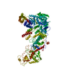 7xw3C M: map data used to model this data C: citing same article ( |
|---|---|
| Similar structure data |
-Links
 PDBj
PDBj


- Assembly
| Deposited unit | 
|
|---|---|
| 1 |
|
-Components
| #1: Protein | Mass: 218947.328 Da / Num. of mol.: 1 Source method: isolated from a genetically manipulated source Source: (gene. exp.) Homo sapiens (human) / Gene: DICER1, DICER, HERNA, KIAA0928 / Production host: Homo sapiens (human) / References: UniProt: Q9UPY3, ribonuclease III | ||
|---|---|---|---|
| #2: RNA chain | Mass: 23343.783 Da / Num. of mol.: 1 / Source method: obtained synthetically / Source: (synth.) Homo sapiens (human) | ||
| #3: Chemical |   Mass: 40.078 Da / Num. of mol.: 2 / Source method: obtained synthetically / Formula: Ca / Feature type: SUBJECT OF INVESTIGATION Mass: 40.078 Da / Num. of mol.: 2 / Source method: obtained synthetically / Formula: Ca / Feature type: SUBJECT OF INVESTIGATIONHas ligand of interest | Y | |
-Experimental details
-Experiment
| Experiment | Method: ELECTRON MICROSCOPY |
|---|---|
| EM experiment | Aggregation state: 3D ARRAY / 3D reconstruction method: single particle reconstruction |
- Sample preparation
| Component |
| ||||||||||||||||||||||||
|---|---|---|---|---|---|---|---|---|---|---|---|---|---|---|---|---|---|---|---|---|---|---|---|---|---|
| Source (natural) | Organism: Homo sapiens (human) | ||||||||||||||||||||||||
| Source (recombinant) | Organism: Homo sapiens (human) | ||||||||||||||||||||||||
| Buffer solution | pH: 8 | ||||||||||||||||||||||||
| Specimen | Embedding applied: NO / Shadowing applied: NO / Staining applied: NO / Vitrification applied: YES | ||||||||||||||||||||||||
| Specimen support | Grid material: GOLD / Grid mesh size: 300 divisions/in. / Grid type: UltrAuFoil R1.2/1.3 | ||||||||||||||||||||||||
| Vitrification | Instrument: FEI VITROBOT MARK IV / Cryogen name: ETHANE / Humidity: 100 % / Chamber temperature: 288 K / Details: blot force 5 and blot for 2 seconds |
- Electron microscopy imaging
| Experimental equipment |  Model: Titan Krios / Image courtesy: FEI Company |
|---|---|
| Microscopy | Model: FEI TITAN KRIOS |
| Electron gun | Electron source:  FIELD EMISSION GUN / Accelerating voltage: 300 kV / Illumination mode: OTHER FIELD EMISSION GUN / Accelerating voltage: 300 kV / Illumination mode: OTHER |
| Electron lens | Mode: BRIGHT FIELD / Nominal defocus max: 2200 nm / Nominal defocus min: 900 nm / Alignment procedure: BASIC |
| Specimen holder | Cryogen: NITROGEN / Specimen holder model: FEI TITAN KRIOS AUTOGRID HOLDER |
| Image recording | Electron dose: 44.163 e/Å2 / Film or detector model: GATAN K3 BIOQUANTUM (6k x 4k) |
- Processing
| CTF correction | Type: NONE |
|---|---|
| 3D reconstruction | Resolution: 3.04 Å / Resolution method: FSC 0.143 CUT-OFF / Num. of particles: 1386301 / Symmetry type: POINT |
