 Movie
Movie Controller


+ Open data

- Basic information
| Entry | Database: PDB / ID: 7xw1 | ||||||
|---|---|---|---|---|---|---|---|
| Title | The crystal structure of AhpD from Pseudomonas aeruginosa | ||||||
 Components Components | Carboxymuconolactone decarboxylase family protein | ||||||
 Keywords Keywords | OXIDOREDUCTASE / AhpD | ||||||
| Function / homology | peroxiredoxin / Alkylhydroperoxidase AhpD core / Carboxymuconolactone decarboxylase-like / Carboxymuconolactone decarboxylase family / AhpD-like / peroxiredoxin activity / Alkyl hydroperoxide reductase AhpD Function and homology information Function and homology information | ||||||
| Biological species |   Pseudomonas aeruginosa (bacteria) Pseudomonas aeruginosa (bacteria) | ||||||
| Method |  X-RAY DIFFRACTION / SYNCHROTRON / MOLECULAR REPLACEMENT / Resolution: 2.36 Å X-RAY DIFFRACTION / SYNCHROTRON / MOLECULAR REPLACEMENT / Resolution: 2.36 Å | ||||||
 Authors Authors | Xu, B. | ||||||
| Funding support | 1items
| ||||||
 Citation Citation | Journal: To Be Published Title: The crystal structure of AhpD from Pseudomonas aeruginosa Authors: Xu, B. | ||||||
| History |
|
- Structure visualization
| Structure viewer | Molecule: MolmilJmol/JSmol |
|---|
- Downloads & links
-Download
| PDBx/mmCIF format | 7xw1.cif.gz | 58.3 KB | Display | PDBx/mmCIF format |
|---|---|---|---|---|
| PDB format | pdb7xw1.ent.gz | 42.1 KB | Display | PDB format |
| PDBx/mmJSON format | 7xw1.json.gz | Tree view | PDBx/mmJSON format | |
| Others |  Other downloads Other downloads |
-Validation report
| Summary document | 7xw1_validation.pdf.gz | 431.6 KB | Display | wwPDB validaton report |
|---|---|---|---|---|
| Full document | 7xw1_full_validation.pdf.gz | 435 KB | Display | |
| Data in XML | 7xw1_validation.xml.gz | 7.2 KB | Display | |
| Data in CIF | 7xw1_validation.cif.gz | 8.5 KB | Display | |
| Arichive directory | https://data.pdbj.org/pub/pdb/validation_reports/xw/7xw1ftp://data.pdbj.org/pub/pdb/validation_reports/xw/7xw1 | HTTPS FTP |
-Related structure data
| Related structure data |  2o4dS S: Starting model for refinement |
|---|---|
| Similar structure data |
-Links
 PDBj
PDBj- Assembly
| Deposited unit | 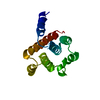
| ||||||||
|---|---|---|---|---|---|---|---|---|---|
| 1 |
| ||||||||
| Unit cell |
|
-Components
| #1: Protein | Mass: 16246.528 Da / Num. of mol.: 1 Source method: isolated from a genetically manipulated source Source: (gene. exp.) Pseudomonas aeruginosa (bacteria) / Production host: |
|---|---|
| Has protein modification | Y |
-Experimental details
-Experiment
| Experiment | Method: X-RAY DIFFRACTION / Number of used crystals: 1 |
|---|
- Sample preparation
| Crystal | Density Matthews: 2.53 Å3/Da / Density % sol: 51.44 % |
|---|---|
| Crystal grow | Temperature: 293 K / Method: vapor diffusion, hanging drop / pH: 7 / Details: 2.4 M Sodium malonate pH 7.0 |
-Data collection
| Diffraction | Mean temperature: 100 K / Serial crystal experiment: N | ||||||||||||||||||||||||||||||
|---|---|---|---|---|---|---|---|---|---|---|---|---|---|---|---|---|---|---|---|---|---|---|---|---|---|---|---|---|---|---|---|
| Diffraction source | Source: SYNCHROTRON / Site: SSRF  / Beamline: BL19U1 / Wavelength: 0.9785 Å / Beamline: BL19U1 / Wavelength: 0.9785 Å | ||||||||||||||||||||||||||||||
| Detector | Type: DECTRIS PILATUS 6M / Detector: PIXEL / Date: Jul 16, 2021 | ||||||||||||||||||||||||||||||
| Radiation | Protocol: SINGLE WAVELENGTH / Monochromatic (M) / Laue (L): M / Scattering type: x-ray | ||||||||||||||||||||||||||||||
| Radiation wavelength | Wavelength: 0.9785 Å / Relative weight: 1 | ||||||||||||||||||||||||||||||
| Reflection | Resolution: 2.36→19.88 Å / Num. obs: 7200 / % possible obs: 98.4 % / Redundancy: 19.8 % / Biso Wilson estimate: 55.63 Å2 / CC1/2: 0.984 / Rmerge(I) obs: 0.27 / Rpim(I) all: 0.062 / Rrim(I) all: 0.277 / Net I/σ(I): 7.8 / Num. measured all: 142387 | ||||||||||||||||||||||||||||||
| Reflection shell | Diffraction-ID: 1
|
- Processing
| Software |
| ||||||||||||||||||||||||||||||||||||||||||
|---|---|---|---|---|---|---|---|---|---|---|---|---|---|---|---|---|---|---|---|---|---|---|---|---|---|---|---|---|---|---|---|---|---|---|---|---|---|---|---|---|---|---|---|
| Refinement | Method to determine structure: MOLECULAR REPLACEMENT Starting model: 2O4D Resolution: 2.36→19.88 Å / SU ML: 0.39 / Cross valid method: THROUGHOUT / σ(F): 1.33 / Phase error: 34.08 / Stereochemistry target values: ML
| ||||||||||||||||||||||||||||||||||||||||||
| Solvent computation | Shrinkage radii: 0.9 Å / VDW probe radii: 1.1 Å / Solvent model: FLAT BULK SOLVENT MODEL | ||||||||||||||||||||||||||||||||||||||||||
| Displacement parameters | Biso max: 149.07 Å2 / Biso mean: 67.1866 Å2 / Biso min: 29.81 Å2 | ||||||||||||||||||||||||||||||||||||||||||
| Refinement step | Cycle: final / Resolution: 2.36→19.88 Å
| ||||||||||||||||||||||||||||||||||||||||||
| LS refinement shell | Refine-ID: X-RAY DIFFRACTION / Rfactor Rfree error: 0 / Total num. of bins used: 5
|
