 Movie
Movie Controller


+ Open data

- Basic information
| Entry | Database: PDB / ID: 7xtr | ||||||||||||
|---|---|---|---|---|---|---|---|---|---|---|---|---|---|
| Title | The apo structure of the engineered TfCut | ||||||||||||
 Components Components | alpha/beta hydrolase | ||||||||||||
 Keywords Keywords | HYDROLASE / PETase / cutinase / enzyme engineering / PBAT degradation | ||||||||||||
| Function / homology | :  Function and homology information Function and homology information | ||||||||||||
| Biological species |   Thermobifida fusca (bacteria) Thermobifida fusca (bacteria) | ||||||||||||
| Method |  X-RAY DIFFRACTION / MOLECULAR REPLACEMENT / Resolution: 2.2 Å X-RAY DIFFRACTION / MOLECULAR REPLACEMENT / Resolution: 2.2 Å | ||||||||||||
 Authors Authors | Yang, Y. / Jiang, P.C. / Huang, J.-W. / Chen, C.-C. / Guo, R.-T. | ||||||||||||
| Funding support |  China, 3items China, 3items
| ||||||||||||
 Citation Citation | Journal: Nat Commun / Year: 2023 Title: Complete bio-degradation of poly(butylene adipate-co-terephthalate) via engineered cutinases. Authors: Yang, Y. / Min, J. / Xue, T. / Jiang, P. / Liu, X. / Peng, R. / Huang, J.W. / Qu, Y. / Li, X. / Ma, N. / Tsai, F.C. / Dai, L. / Zhang, Q. / Liu, Y. / Chen, C.C. / Guo, R.T. | ||||||||||||
| History |
|
- Structure visualization
| Structure viewer | Molecule: MolmilJmol/JSmol |
|---|
- Downloads & links
-Download
| PDBx/mmCIF format | 7xtr.cif.gz | 118.2 KB | Display | PDBx/mmCIF format |
|---|---|---|---|---|
| PDB format | pdb7xtr.ent.gz | 89.2 KB | Display | PDB format |
| PDBx/mmJSON format | 7xtr.json.gz | Tree view | PDBx/mmJSON format | |
| Others |  Other downloads Other downloads |
-Validation report
| Arichive directory | https://data.pdbj.org/pub/pdb/validation_reports/xt/7xtrftp://data.pdbj.org/pub/pdb/validation_reports/xt/7xtr | HTTPS FTP |
|---|
-Related structure data
| Related structure data | 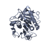 7xtsC 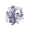 7xttC 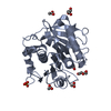 7xtuC  7xtvC  7xtwC 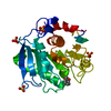 4cg2S S: Starting model for refinement C: citing same article ( |
|---|---|
| Similar structure data |
-Links
 PDBj
PDBj
- Assembly
| Deposited unit | 
| ||||||||
|---|---|---|---|---|---|---|---|---|---|
| 1 | 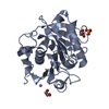
| ||||||||
| 2 | 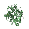
| ||||||||
| Unit cell |
|
-Components
| #1: Protein | Mass: 28233.678 Da / Num. of mol.: 2 / Mutation: H184S,F188I Source method: isolated from a genetically manipulated source Details: GB:PZN61876.1 / Source: (gene. exp.) Thermobifida fusca (bacteria) / Gene: cut-2.KW3 / Production host: #2: Chemical |   Mass: 96.063 Da / Num. of mol.: 2 / Source method: obtained synthetically / Formula: SO4 Mass: 96.063 Da / Num. of mol.: 2 / Source method: obtained synthetically / Formula: SO4#3: Chemical |   Mass: 92.094 Da / Num. of mol.: 2 / Source method: obtained synthetically / Formula: C3H8O3 Mass: 92.094 Da / Num. of mol.: 2 / Source method: obtained synthetically / Formula: C3H8O3#4: Chemical | ChemComp-LI / |   Mass: 6.941 Da / Num. of mol.: 1 / Source method: obtained synthetically / Formula: Li Mass: 6.941 Da / Num. of mol.: 1 / Source method: obtained synthetically / Formula: Li#5: Water | ChemComp-HOH / |  Mass: 18.015 Da / Num. of mol.: 338 / Source method: isolated from a natural source / Formula: H2O Mass: 18.015 Da / Num. of mol.: 338 / Source method: isolated from a natural source / Formula: H2OHas ligand of interest | N | Has protein modification | Y | |
|---|
-Experimental details
-Experiment
| Experiment | Method: X-RAY DIFFRACTION / Number of used crystals: 1 |
|---|
- Sample preparation
| Crystal | Density Matthews: 1.97 Å3/Da / Density % sol: 37.54 % |
|---|---|
| Crystal grow | Temperature: 273 K / Method: evaporation / pH: 8.5 Details: 20% w/v PEG3350, 0.2 M Sodium nitrate, 0.1 M BIS-Tris propane 8.5 |
-Data collection
| Diffraction | Mean temperature: 100 K / Serial crystal experiment: N |
|---|---|
| Diffraction source | Source: LIQUID ANODE / Type: BRUKER METALJET / Wavelength: 1.34138 Å |
| Detector | Type: BRUKER PHOTON 100 / Detector: CMOS / Date: Nov 21, 2020 |
| Radiation | Protocol: SINGLE WAVELENGTH / Monochromatic (M) / Laue (L): M / Scattering type: x-ray |
| Radiation wavelength | Wavelength: 1.34138 Å / Relative weight: 1 |
| Reflection | Resolution: 2.2→37.2 Å / Num. obs: 38580 / % possible obs: 96.5 % / Redundancy: 4.2 % / Rmerge(I) obs: 0.064 / Net I/σ(I): 16.1 |
| Reflection shell | Resolution: 2.2→2.23 Å / Rmerge(I) obs: 0.099 / Mean I/σ(I) obs: 3 / Num. unique obs: 860 |
- Processing
| Software |
| ||||||||||||||||||||||||||||||||||||||||||||||||||||||||||||||||||||||||||||||||||||||||||||||||||||||||||||||||||||||||||||||||||||||||||||||||||||||||||||||||||||||||||||||||||||||
|---|---|---|---|---|---|---|---|---|---|---|---|---|---|---|---|---|---|---|---|---|---|---|---|---|---|---|---|---|---|---|---|---|---|---|---|---|---|---|---|---|---|---|---|---|---|---|---|---|---|---|---|---|---|---|---|---|---|---|---|---|---|---|---|---|---|---|---|---|---|---|---|---|---|---|---|---|---|---|---|---|---|---|---|---|---|---|---|---|---|---|---|---|---|---|---|---|---|---|---|---|---|---|---|---|---|---|---|---|---|---|---|---|---|---|---|---|---|---|---|---|---|---|---|---|---|---|---|---|---|---|---|---|---|---|---|---|---|---|---|---|---|---|---|---|---|---|---|---|---|---|---|---|---|---|---|---|---|---|---|---|---|---|---|---|---|---|---|---|---|---|---|---|---|---|---|---|---|---|---|---|---|---|---|
| Refinement | Method to determine structure: MOLECULAR REPLACEMENT Starting model: 4CG2 Resolution: 2.2→32.68 Å / SU ML: 0.25 / Cross valid method: THROUGHOUT / σ(F): 1.35 / Phase error: 22.74 / Stereochemistry target values: ML
| ||||||||||||||||||||||||||||||||||||||||||||||||||||||||||||||||||||||||||||||||||||||||||||||||||||||||||||||||||||||||||||||||||||||||||||||||||||||||||||||||||||||||||||||||||||||
| Solvent computation | Shrinkage radii: 0.9 Å / VDW probe radii: 1.11 Å / Solvent model: FLAT BULK SOLVENT MODEL | ||||||||||||||||||||||||||||||||||||||||||||||||||||||||||||||||||||||||||||||||||||||||||||||||||||||||||||||||||||||||||||||||||||||||||||||||||||||||||||||||||||||||||||||||||||||
| Displacement parameters | Biso max: 62.45 Å2 / Biso mean: 16.2891 Å2 / Biso min: 3.78 Å2 | ||||||||||||||||||||||||||||||||||||||||||||||||||||||||||||||||||||||||||||||||||||||||||||||||||||||||||||||||||||||||||||||||||||||||||||||||||||||||||||||||||||||||||||||||||||||
| Refinement step | Cycle: final / Resolution: 2.2→32.68 Å
| ||||||||||||||||||||||||||||||||||||||||||||||||||||||||||||||||||||||||||||||||||||||||||||||||||||||||||||||||||||||||||||||||||||||||||||||||||||||||||||||||||||||||||||||||||||||
| LS refinement shell | Refine-ID: X-RAY DIFFRACTION / Rfactor Rfree error: 0 / Total num. of bins used: 25
|
