 Movie
Movie Controller


+ Open data

- Basic information
| Entry | Database: PDB / ID: 7xrm | ||||||
|---|---|---|---|---|---|---|---|
| Title | Ethanolamine ammonia-lyase complexed with AdoMeCbl | ||||||
 Components Components | (Ethanolamine ammonia-lyase ...) x 2 | ||||||
 Keywords Keywords | LYASE / Adenosylcobalamin / Radical enzyme | ||||||
| Function / homology |  Function and homology information Function and homology informationethanolamine ammonia-lyase / ethanolamine ammonia-lyase activity / ethanolamine ammonia-lyase complex / ethanolamine degradation polyhedral organelle / ethanolamine catabolic process / cobalamin binding / amino acid metabolic process / cytosol Similarity search - Function | ||||||
| Biological species |  | ||||||
| Method |  X-RAY DIFFRACTION / SYNCHROTRON / MOLECULAR REPLACEMENT / Resolution: 2.13 Å X-RAY DIFFRACTION / SYNCHROTRON / MOLECULAR REPLACEMENT / Resolution: 2.13 Å | ||||||
 Authors Authors | Shibata, N. / Toraya, T. | ||||||
| Funding support | 1items
| ||||||
 Citation Citation | Journal: Chemistry / Year: 2022 Title: Structural Insights into the Very Low Activity of the Homocoenzyme B 12 Adenosylmethylcobalamin in Coenzyme B 12 -Dependent Diol Dehydratase and Ethanolamine Ammonia-Lyase. Authors: Shibata, N. / Higuchi, Y. / Krautler, B. / Toraya, T. | ||||||
| History |
|
- Structure visualization
| Structure viewer | Molecule: MolmilJmol/JSmol |
|---|
- Downloads & links
-Download
| PDBx/mmCIF format | 7xrm.cif.gz | 693.1 KB | Display | PDBx/mmCIF format |
|---|---|---|---|---|
| PDB format | pdb7xrm.ent.gz | 473.3 KB | Display | PDB format |
| PDBx/mmJSON format | 7xrm.json.gz | Tree view | PDBx/mmJSON format | |
| Others |  Other downloads Other downloads |
-Validation report
| Arichive directory | https://data.pdbj.org/pub/pdb/validation_reports/xr/7xrmftp://data.pdbj.org/pub/pdb/validation_reports/xr/7xrm | HTTPS FTP |
|---|
-Related structure data
| Related structure data |  7xrkC  7xrlC  7xrnC  3abrS S: Starting model for refinement C: citing same article ( |
|---|---|
| Similar structure data |
-Links
 PDBj
PDBj
- Assembly
| Deposited unit | 
| ||||||||||||
|---|---|---|---|---|---|---|---|---|---|---|---|---|---|
| 1 | 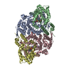
| ||||||||||||
| Unit cell |
|
-Components
-Ethanolamine ammonia-lyase ... , 2 types, 4 molecules ACBD
| #1: Protein | Mass: 49447.816 Da / Num. of mol.: 2 Source method: isolated from a genetically manipulated source Source: (gene. exp.) #2: Protein | Mass: 28769.021 Da / Num. of mol.: 2 Source method: isolated from a genetically manipulated source Source: (gene. exp.) |
|---|
-Non-polymers , 5 types, 668 molecules 


| #3: Chemical | ChemComp-GOL /  Mass: 92.094 Da / Num. of mol.: 11 / Source method: obtained synthetically / Formula: C3H8O3 Mass: 92.094 Da / Num. of mol.: 11 / Source method: obtained synthetically / Formula: C3H8O3#4: Chemical |  Mass: 18.038 Da / Num. of mol.: 2 / Source method: obtained synthetically / Formula: H4N Mass: 18.038 Da / Num. of mol.: 2 / Source method: obtained synthetically / Formula: H4N#5: Chemical |  Mass: 265.269 Da / Num. of mol.: 2 / Source method: obtained synthetically / Formula: C11H15N5O3 / Feature type: SUBJECT OF INVESTIGATION Mass: 265.269 Da / Num. of mol.: 2 / Source method: obtained synthetically / Formula: C11H15N5O3 / Feature type: SUBJECT OF INVESTIGATION#6: Chemical |  Mass: 1330.356 Da / Num. of mol.: 2 / Source method: obtained synthetically / Formula: C62H89CoN13O14P / Feature type: SUBJECT OF INVESTIGATION Mass: 1330.356 Da / Num. of mol.: 2 / Source method: obtained synthetically / Formula: C62H89CoN13O14P / Feature type: SUBJECT OF INVESTIGATION#7: Water | ChemComp-HOH / | Mass: 18.015 Da / Num. of mol.: 651 / Source method: isolated from a natural source / Formula: H2O |
|---|
-Details
| Has ligand of interest | Y |
|---|
-Experimental details
-Experiment
| Experiment | Method: X-RAY DIFFRACTION / Number of used crystals: 1 |
|---|
- Sample preparation
| Crystal | Density Matthews: 4.17 Å3/Da / Density % sol: 70.51 % |
|---|---|
| Crystal grow | Temperature: 277 K / Method: vapor diffusion, sitting drop / pH: 7 Details: 10% (w/v) PEG 6000, 0.1 M ammonium chloride, and 0.05 M HEPES pH 7.0 |
-Data collection
| Diffraction | Mean temperature: 100 K / Serial crystal experiment: N |
|---|---|
| Diffraction source | Source: SYNCHROTRON / Site: SPring-8  / Beamline: BL44XU / Wavelength: 0.9 Å / Beamline: BL44XU / Wavelength: 0.9 Å |
| Detector | Type: RAYONIX MX300HE / Detector: CCD / Date: Feb 10, 2015 |
| Radiation | Protocol: SINGLE WAVELENGTH / Monochromatic (M) / Laue (L): M / Scattering type: x-ray |
| Radiation wavelength | Wavelength: 0.9 Å / Relative weight: 1 |
| Reflection | Resolution: 2.13→47 Å / Num. obs: 143007 / % possible obs: 99.2 % / Redundancy: 7.71 % / CC1/2: 0.998 / Rmerge(I) obs: 0.123 / Rrim(I) all: 0.132 / Net I/σ(I): 14.95 |
| Reflection shell | Resolution: 2.13→2.19 Å / Redundancy: 7.64 % / Rmerge(I) obs: 1.173 / Num. unique obs: 10603 / CC1/2: 0.479 / Rrim(I) all: 1.258 / % possible all: 99.8 |
- Processing
| Software |
| |||||||||||||||||||||||||||||||||||||||||||||||||||||||||||||||||||||||||||||||||||||||||||||||||||||||||||||||||||||||||||||||||||||||||||||||||||
|---|---|---|---|---|---|---|---|---|---|---|---|---|---|---|---|---|---|---|---|---|---|---|---|---|---|---|---|---|---|---|---|---|---|---|---|---|---|---|---|---|---|---|---|---|---|---|---|---|---|---|---|---|---|---|---|---|---|---|---|---|---|---|---|---|---|---|---|---|---|---|---|---|---|---|---|---|---|---|---|---|---|---|---|---|---|---|---|---|---|---|---|---|---|---|---|---|---|---|---|---|---|---|---|---|---|---|---|---|---|---|---|---|---|---|---|---|---|---|---|---|---|---|---|---|---|---|---|---|---|---|---|---|---|---|---|---|---|---|---|---|---|---|---|---|---|---|---|---|
| Refinement | Method to determine structure: MOLECULAR REPLACEMENT Starting model: 3ABR Resolution: 2.13→46.42 Å / Cross valid method: FREE R-VALUE / σ(F): 58.54 / Phase error: 26.0603 Stereochemistry target values: GeoStd + Monomer Library + CDL v1.2
| |||||||||||||||||||||||||||||||||||||||||||||||||||||||||||||||||||||||||||||||||||||||||||||||||||||||||||||||||||||||||||||||||||||||||||||||||||
| Solvent computation | Shrinkage radii: 0.9 Å / VDW probe radii: 1.11 Å / Solvent model: FLAT BULK SOLVENT MODEL | |||||||||||||||||||||||||||||||||||||||||||||||||||||||||||||||||||||||||||||||||||||||||||||||||||||||||||||||||||||||||||||||||||||||||||||||||||
| Displacement parameters | Biso mean: 47.99 Å2 | |||||||||||||||||||||||||||||||||||||||||||||||||||||||||||||||||||||||||||||||||||||||||||||||||||||||||||||||||||||||||||||||||||||||||||||||||||
| Refinement step | Cycle: LAST / Resolution: 2.13→46.42 Å
| |||||||||||||||||||||||||||||||||||||||||||||||||||||||||||||||||||||||||||||||||||||||||||||||||||||||||||||||||||||||||||||||||||||||||||||||||||
| Refine LS restraints |
| |||||||||||||||||||||||||||||||||||||||||||||||||||||||||||||||||||||||||||||||||||||||||||||||||||||||||||||||||||||||||||||||||||||||||||||||||||
| LS refinement shell |
| |||||||||||||||||||||||||||||||||||||||||||||||||||||||||||||||||||||||||||||||||||||||||||||||||||||||||||||||||||||||||||||||||||||||||||||||||||
| Refinement TLS params. | Method: refined / Refine-ID: X-RAY DIFFRACTION
| |||||||||||||||||||||||||||||||||||||||||||||||||||||||||||||||||||||||||||||||||||||||||||||||||||||||||||||||||||||||||||||||||||||||||||||||||||
| Refinement TLS group | Refine-ID: X-RAY DIFFRACTION
|
