 Movie
Movie Controller

[English] 日本語
 Yorodumi
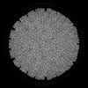
- PDB-7xr2: 3.1 Angstrom cryoEM icosahedral reconstruction of mud crab reovirus -
+ Open data

- Basic information
| Entry | Database: PDB / ID: 7xr2 | ||||||||||||||||||||||||||||||||||||||||||||||||||||||
|---|---|---|---|---|---|---|---|---|---|---|---|---|---|---|---|---|---|---|---|---|---|---|---|---|---|---|---|---|---|---|---|---|---|---|---|---|---|---|---|---|---|---|---|---|---|---|---|---|---|---|---|---|---|---|---|
| Title | 3.1 Angstrom cryoEM icosahedral reconstruction of mud crab reovirus | ||||||||||||||||||||||||||||||||||||||||||||||||||||||
 Components Components |
| ||||||||||||||||||||||||||||||||||||||||||||||||||||||
 Keywords Keywords | VIRUS / Reovirus / ds-RNA virus | ||||||||||||||||||||||||||||||||||||||||||||||||||||||
| Function / homology | VP3 / VP11 / VP12 Function and homology information Function and homology information | ||||||||||||||||||||||||||||||||||||||||||||||||||||||
| Biological species |  Scylla serrata reovirus SZ-2007 Scylla serrata reovirus SZ-2007 | ||||||||||||||||||||||||||||||||||||||||||||||||||||||
| Method | ELECTRON MICROSCOPY / single particle reconstruction / cryo EM / Resolution: 3.1 Å | ||||||||||||||||||||||||||||||||||||||||||||||||||||||
 Authors Authors | Zhang, Q. / Gao, Y. | ||||||||||||||||||||||||||||||||||||||||||||||||||||||
| Funding support |  China, 2items China, 2items
| ||||||||||||||||||||||||||||||||||||||||||||||||||||||
 Citation Citation | Journal: PLoS Pathog / Year: 2023 Title: The structure of a 12-segmented dsRNA reovirus: New insights into capsid stabilization and organization. Authors: Qinfen Zhang / Yuanzhu Gao / Matthew L Baker / Shanshan Liu / Xudong Jia / Haidong Xu / Jianguo He / Jason T Kaelber / Shaoping Weng / Wen Jiang /  Abstract: Infecting a wide range of hosts, members of Reovirales (formerly Reoviridae) consist of a genome with different numbers of segmented double stranded RNAs (dsRNA) encapsulated by a proteinaceous shell ...Infecting a wide range of hosts, members of Reovirales (formerly Reoviridae) consist of a genome with different numbers of segmented double stranded RNAs (dsRNA) encapsulated by a proteinaceous shell and carry out genome replication and transcription inside the virion. Several cryo-electron microscopy (cryo-EM) structures of reoviruses with 9, 10 or 11 segmented dsRNA genomes have revealed insights into genome arrangement and transcription. However, the structure and genome arrangement of 12-segmented Reovirales members remain poorly understood. Using cryo-EM, we determined the structure of mud crab reovirus (MCRV), a 12-segmented dsRNA virus that is a putative member of Reovirales in the non-turreted Sedoreoviridae family, to near-atomic resolutions with icosahedral symmetry (3.1 Å) and without imposing icosahedral symmetry (3.4 Å). These structures revealed the organization of the major capsid proteins in two layers: an outer T = 13 layer consisting of VP12 trimers and unique VP11 clamps, and an inner T = 1 layer consisting of VP3 dimers. Additionally, ten RNA dependent RNA polymerases (RdRp) were well resolved just below the VP3 layer but were offset from the 5-fold axes and arranged with D5 symmetry, which has not previously been seen in other members of Reovirales. The N-termini of VP3 were shown to adopt four unique conformations; two of which anchor the RdRps, while the other two conformations are likely involved in genome organization and capsid stability. Taken together, these structures provide a new level of understanding for capsid stabilization and genome organization of segmented dsRNA viruses. | ||||||||||||||||||||||||||||||||||||||||||||||||||||||
| History |
|
- Structure visualization
| Structure viewer | Molecule: MolmilJmol/JSmol |
|---|
- Downloads & links
-Download
| PDBx/mmCIF format | 7xr2.cif.gz | 924.8 KB | Display | PDBx/mmCIF format |
|---|---|---|---|---|
| PDB format | pdb7xr2.ent.gz | 783.2 KB | Display | PDB format |
| PDBx/mmJSON format | 7xr2.json.gz | Tree view | PDBx/mmJSON format | |
| Others |  Other downloads Other downloads |
-Validation report
| Arichive directory | https://data.pdbj.org/pub/pdb/validation_reports/xr/7xr2ftp://data.pdbj.org/pub/pdb/validation_reports/xr/7xr2 | HTTPS FTP |
|---|
-Related structure data
| Related structure data | 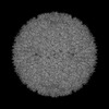 33403MC  7xr3C M: map data used to model this data C: citing same article ( |
|---|---|
| Similar structure data |


-Links
 PDBj
PDBj- Assembly
| Deposited unit | 
|
|---|---|
| 1 | x 60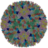
|
| 2 |
|
| 3 | x 5
|
| 4 | x 6
|
| 5 | 
|
| Symmetry | Point symmetry: (Schoenflies symbol: I (icosahedral)) |
-Components
| #1: Protein | Mass: 97019.383 Da / Num. of mol.: 2 / Source method: isolated from a natural source / Source: (natural) Scylla serrata reovirus SZ-2007 / References: UniProt: E9LEU6#2: Protein | Mass: 23750.701 Da / Num. of mol.: 2 / Source method: isolated from a natural source / Source: (natural) Scylla serrata reovirus SZ-2007 / References: UniProt: G9BDA7#3: Protein | Mass: 29730.963 Da / Num. of mol.: 13 / Source method: isolated from a natural source / Source: (natural) Scylla serrata reovirus SZ-2007 / References: UniProt: G9BDA8Has protein modification | N | |
|---|
-Experimental details
-Experiment
| Experiment | Method: ELECTRON MICROSCOPY |
|---|---|
| EM experiment | Aggregation state: PARTICLE / 3D reconstruction method: single particle reconstruction |
- Sample preparation
| Component | Name: Scylla serrata reovirus SZ-2007 / Type: VIRUS / Entity ID: all / Source: NATURAL |
|---|---|
| Source (natural) | Organism: Scylla serrata reovirus SZ-2007 |
| Details of virus | Empty: NO / Enveloped: NO / Isolate: STRAIN / Type: VIRION |
| Natural host | Organism: Scylla serrata |
| Buffer solution | pH: 7.4 |
| Specimen | Embedding applied: NO / Shadowing applied: NO / Staining applied: NO / Vitrification applied: YES |
| Specimen support | Grid material: COPPER / Grid mesh size: 300 divisions/in. / Grid type: Quantifoil R1.2/1.3 |
| Vitrification | Instrument: FEI VITROBOT MARK IV / Cryogen name: ETHANE / Humidity: 100 % / Chamber temperature: 277 K |
- Electron microscopy imaging
| Experimental equipment |  Model: Titan Krios / Image courtesy: FEI Company |
|---|---|
| Microscopy | Model: FEI TITAN KRIOS |
| Electron gun | Electron source:  FIELD EMISSION GUN / Accelerating voltage: 300 kV / Illumination mode: FLOOD BEAM FIELD EMISSION GUN / Accelerating voltage: 300 kV / Illumination mode: FLOOD BEAM |
| Electron lens | Mode: BRIGHT FIELD / Nominal magnification: 75000 X / Calibrated magnification: 128440 X / Nominal defocus max: 3000 nm / Nominal defocus min: 600 nm / Calibrated defocus min: 600 nm / Calibrated defocus max: 3000 nm / Cs: 2.7 mm / C2 aperture diameter: 70 µm / Alignment procedure: COMA FREE |
| Specimen holder | Cryogen: NITROGEN / Specimen holder model: FEI TITAN KRIOS AUTOGRID HOLDER / Temperature (max): 100 K / Temperature (min): 80 K |
| Image recording | Average exposure time: 1.1 sec. / Electron dose: 25 e/Å2 / Detector mode: INTEGRATING / Film or detector model: FEI FALCON II (4k x 4k) / Num. of grids imaged: 1 / Num. of real images: 3595 |
| Image scans | Sampling size: 14 µm / Width: 4096 / Height: 4096 / Movie frames/image: 16 / Used frames/image: 1-16 |
- Processing
| Software | Name: PHENIX / Version: 1.14rc1_3161: / Classification: refinement | ||||||||||||||||||||||||||||||||
|---|---|---|---|---|---|---|---|---|---|---|---|---|---|---|---|---|---|---|---|---|---|---|---|---|---|---|---|---|---|---|---|---|---|
| EM software |
| ||||||||||||||||||||||||||||||||
| CTF correction | Type: NONE | ||||||||||||||||||||||||||||||||
| Particle selection | Num. of particles selected: 58095 | ||||||||||||||||||||||||||||||||
| 3D reconstruction | Resolution: 3.1 Å / Resolution method: FSC 0.143 CUT-OFF / Num. of particles: 58095 / Algorithm: FOURIER SPACE / Symmetry type: POINT |
