- PDB-7xlo: Crystal Structure of the Catalytic Domain of Inosine Monophosphat... -
+
Open data
ID or keywords:
Loading...
-
Basic information
Entry
Database: PDB / ID: 7xlo
Title
Crystal Structure of the Catalytic Domain of Inosine Monophosphate Dehydrogenase (IMPDH) from Methanocaldococcus jannaschii
Components
Inosine-5'-monophosphate dehydrogenase
Keywords
OXIDOREDUCTASE / IMPDH TIM / Catalytic domain / alpha and beta proteins (a/b) / TIM beta/alpha-barrel
Function / homology
Function and homology information
IMP dehydrogenase / IMP dehydrogenase activity / GMP biosynthetic process / GTP biosynthetic process / nucleotide binding / metal ion binding Similarity search - Function
IMP dehydrogenase / GMP reductase, conserved site / IMP dehydrogenase / GMP reductase signature. / Inosine-5'-monophosphate dehydrogenase / IMP dehydrogenase/GMP reductase / IMP dehydrogenase / GMP reductase domain / IMP dehydrogenase / GMP reductase domain / Domain in cystathionine beta-synthase and other proteins. / CBS domain superfamily / CBS domain / CBS domain ...IMP dehydrogenase / GMP reductase, conserved site / IMP dehydrogenase / GMP reductase signature. / Inosine-5'-monophosphate dehydrogenase / IMP dehydrogenase/GMP reductase / IMP dehydrogenase / GMP reductase domain / IMP dehydrogenase / GMP reductase domain / Domain in cystathionine beta-synthase and other proteins. / CBS domain superfamily / CBS domain / CBS domain / CBS domain profile. / Aldolase class I / Aldolase-type TIM barrel / TIM Barrel / Alpha-Beta Barrel / Alpha Beta Similarity search - Domain/homology
Resolution: 2.6→53.37 Å / Cor.coef. Fo:Fc: 0.827 / Cor.coef. Fo:Fc free: 0.806 / SU B: 15.822 / SU ML: 0.309 / Cross valid method: THROUGHOUT / ESU R: 0.843 / ESU R Free: 0.339 / Stereochemistry target values: MAXIMUM LIKELIHOOD / Details: HYDROGENS HAVE BEEN ADDED IN THE RIDING POSITIONS
Rfactor
Num. reflection
% reflection
Selection details
Rfree
0.27606
1233
5.1 %
RANDOM
Rwork
0.25416
-
-
-
obs
0.25525
22945
99.98 %
-
Solvent computation
Ion probe radii: 0.8 Å / Shrinkage radii: 0.8 Å / VDW probe radii: 1.2 Å / Solvent model: MASK
 Movie
Movie Controller

 Yorodumi
 Open data
 Basic information
 Components
 Keywords
 Function and homology information
 Methanocaldococcus jannaschii (archaea)
 X-RAY DIFFRACTION /
  Authors
 India, 2items
  Citation
 Structure visualization
 Downloads & links
 Other downloads
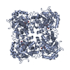
 PDBj
PDBj
 Assembly


 Mass: 18.015 Da / Num. of mol.: 165 / Source method: isolated from a natural source / Formula: H2O
 Sample preparation
 Processing