 Movie
Movie Controller


+ Open data

- Basic information
| Entry | Database: PDB / ID: 7xkj | ||||||
|---|---|---|---|---|---|---|---|
| Title | Kras-G12D-GDP-MRTX1133 by FIB-MicroED | ||||||
 Components Components | KRAS proto-oncogene, GTPase | ||||||
 Keywords Keywords | ONCOPROTEIN / g12d / gdp / gtpase / hydrolase-hydrolase inhibitor complex / hydrolase/hydrolase inhibitor / MicroED | ||||||
| Function / homology |  Function and homology information Function and homology informationsmall monomeric GTPase / GDP binding / Ras protein signal transduction / GTPase activity / GTP binding / plasma membrane Similarity search - Function | ||||||
| Biological species |  Homo sapiens (human) Homo sapiens (human) | ||||||
| Method | ELECTRON CRYSTALLOGRAPHY / electron crystallography / cryo EM / Resolution: 3 Å | ||||||
 Authors Authors | Li, X.M. | ||||||
| Funding support |  China, 1items China, 1items
| ||||||
 Citation Citation | Journal: To Be Published Title: Kras-G12D-GDP-MRTX1133 by FIB-MicroED Authors: Li, X.M. | ||||||
| History |
|
- Structure visualization
| Structure viewer | Molecule: MolmilJmol/JSmol |
|---|
- Downloads & links
-Download
| PDBx/mmCIF format | 7xkj.cif.gz | 58.9 KB | Display | PDBx/mmCIF format |
|---|---|---|---|---|
| PDB format | pdb7xkj.ent.gz | 33.7 KB | Display | PDB format |
| PDBx/mmJSON format | 7xkj.json.gz | Tree view | PDBx/mmJSON format | |
| Others |  Other downloads Other downloads |
-Validation report
| Arichive directory | https://data.pdbj.org/pub/pdb/validation_reports/xk/7xkjftp://data.pdbj.org/pub/pdb/validation_reports/xk/7xkj | HTTPS FTP |
|---|
-Related structure data
| Similar structure data |
|---|
-Links
 PDBj
PDBj


- Assembly
| Deposited unit | 
| ||||||||||||
|---|---|---|---|---|---|---|---|---|---|---|---|---|---|
| 1 |
| ||||||||||||
| Unit cell |
|
 gel filtration
-Components
| #1: Protein | Mass: 19313.730 Da / Num. of mol.: 1 / Mutation: G12D, C118S Source method: isolated from a genetically manipulated source Source: (gene. exp.) Homo sapiens (human) / Gene: mMyoMyo1_007468 / Production host:  |
|---|---|
| #2: Chemical | ChemComp-GDP /   Type: RNA linking / Mass: 443.201 Da / Num. of mol.: 1 / Source method: obtained synthetically / Formula: C10H15N5O11P2 / Feature type: SUBJECT OF INVESTIGATION / Comment: GDP, energy-carrying molecule*YM Type: RNA linking / Mass: 443.201 Da / Num. of mol.: 1 / Source method: obtained synthetically / Formula: C10H15N5O11P2 / Feature type: SUBJECT OF INVESTIGATION / Comment: GDP, energy-carrying molecule*YM |
| #3: Chemical | ChemComp-6IC / 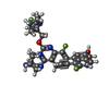  Mass: 600.633 Da / Num. of mol.: 1 / Source method: obtained synthetically / Formula: C33H31F3N6O2 / Feature type: SUBJECT OF INVESTIGATION Mass: 600.633 Da / Num. of mol.: 1 / Source method: obtained synthetically / Formula: C33H31F3N6O2 / Feature type: SUBJECT OF INVESTIGATION |
| #4: Chemical | ChemComp-MG /   Mass: 24.305 Da / Num. of mol.: 1 / Source method: obtained synthetically / Formula: Mg / Feature type: SUBJECT OF INVESTIGATION Mass: 24.305 Da / Num. of mol.: 1 / Source method: obtained synthetically / Formula: Mg / Feature type: SUBJECT OF INVESTIGATION |
| #5: Water | ChemComp-HOH /  Mass: 18.015 Da / Num. of mol.: 6 / Source method: isolated from a natural source / Formula: H2O Mass: 18.015 Da / Num. of mol.: 6 / Source method: isolated from a natural source / Formula: H2O |
| Has ligand of interest | Y |
| Has protein modification | N |
-Experimental details
-Experiment
| Experiment | Method: ELECTRON CRYSTALLOGRAPHY |
|---|---|
| EM experiment | Aggregation state: 3D ARRAY / 3D reconstruction method: electron crystallography |
- Sample preparation
| Component | Name: Isoform 2B of GTPase KRas GUANOSINE-5'-DIPHOSPHATE 4-(4-[(1R,5S)-3,8-diazabicyclo[3.2.1]octan-3-yl]-8-fluoro-2-{[(2R,4R,7aS)-2-fluorotetrahydro-1H-pyrrolizin-7a(5H)-yl]methoxy}pyrido[4,3-d] ...Name: Isoform 2B of GTPase KRas GUANOSINE-5'-DIPHOSPHATE 4-(4-[(1R,5S)-3,8-diazabicyclo[3.2.1]octan-3-yl]-8-fluoro-2-{[(2R,4R,7aS)-2-fluorotetrahydro-1H-pyrrolizin-7a(5H)-yl]methoxy}pyrido[4,3-d]pyrimidin-7-yl)-5-ethynyl-6-fluoronaphthalen-2-ol MAGNESIUM ION Type: COMPLEX / Entity ID: #1 / Source: RECOMBINANT |
|---|---|
| Molecular weight | Value: 0.02 MDa / Experimental value: YES |
| Source (natural) | Organism: Homo sapiens (human) |
| Source (recombinant) | Organism: |
| Buffer solution | pH: 8 |
| Specimen | Embedding applied: NO / Shadowing applied: NO / Staining applied: NO / Vitrification applied: YES |
| Vitrification | Cryogen name: ETHANE |
-Data collection
| Microscopy | Model: JEOL 2100F |
|---|---|
| Electron gun | Electron source: LAB6 / Accelerating voltage: 200 kV / Illumination mode: FLOOD BEAM |
| Electron lens | Mode: DIFFRACTION / Nominal magnification: 6000 X / Calibrated magnification: 6000 X / Nominal defocus max: 2500 nm / Nominal defocus min: 1500 nm / Calibrated defocus min: 1200 nm / Calibrated defocus max: 2300 nm / C2 aperture diameter: 100 µm / Alignment procedure: BASIC |
| Specimen holder | Cryogen: NITROGEN / Specimen holder model: FISCHIONE 2550 / Temperature (max): 100 K / Temperature (min): 90 K |
| Image recording | Average exposure time: 2 sec. / Electron dose: 0.02 e/Å2 / Detector mode: OTHER / Film or detector model: DIRECT ELECTRON DE-20 (5k x 3k) |
| EM imaging optics | Phase plate: OTHER |
| EM diffraction | Camera length: 100 mm |
| EM diffraction shell | Resolution: 3→33.17 Å / Fourier space coverage: 100 % / Multiplicity: 8.3 / Num. of structure factors: 3591 / Phase residual: 13.5 ° |
| EM diffraction stats | Fourier space coverage: 100 % / High resolution: 3 Å / Num. of intensities measured: 29866 / Num. of structure factors: 3591 / Phase error rejection criteria: no / Rmerge: 0.4827 |
| Diffraction source | Wavelength: 0.025 |
| Radiation wavelength | Wavelength: 0.025 Å / Relative weight: 1 |
| Reflection | Biso Wilson estimate: 44.23 Å2 |
- Processing
| Software | Name: PHENIX / Version: 1.19.2_4158 / Classification: refinement / Contact author: Paul D. Adams / Contact author email: pdadams[at]lbl.gov / Language: Python/C++ / URL: https://www.phenix-online.org/ / Type: program | ||||||||||||||||||||||||||||||||||||||||||
|---|---|---|---|---|---|---|---|---|---|---|---|---|---|---|---|---|---|---|---|---|---|---|---|---|---|---|---|---|---|---|---|---|---|---|---|---|---|---|---|---|---|---|---|
| EM software |
| ||||||||||||||||||||||||||||||||||||||||||
| EM 3D crystal entity | ∠α: 90 ° / ∠β: 90 ° / ∠γ: 90 ° / A: 39.868 Å / B: 51.757 Å / C: 89.611 Å / Space group name: P212121 / Space group num: 19 | ||||||||||||||||||||||||||||||||||||||||||
| CTF correction | Type: NONE | ||||||||||||||||||||||||||||||||||||||||||
| 3D reconstruction | Method: CRYSTALLOGRAPHY / Resolution method: DIFFRACTION PATTERN/LAYERLINES / Symmetry type: 3D CRYSTAL | ||||||||||||||||||||||||||||||||||||||||||
| Atomic model building | B value: 200 / Protocol: OTHER / Space: REAL / Target criteria: Correlation coefficient / Details: no | ||||||||||||||||||||||||||||||||||||||||||
| Atomic model building | PDB-ID: 7RPZ Pdb chain-ID: A / Accession code: 7RPZ / Pdb chain residue range: 1-169 / Source name: PDB / Type: experimental model | ||||||||||||||||||||||||||||||||||||||||||
| Refinement | Resolution: 3→35.51 Å / SU ML: 0.3835 / Cross valid method: FREE R-VALUE / σ(F): 1.35 / Phase error: 31.2454 Stereochemistry target values: GeoStd + Monomer Library + CDL v1.2
| ||||||||||||||||||||||||||||||||||||||||||
| Solvent computation | Shrinkage radii: 0.9 Å / VDW probe radii: 1.11 Å / Solvent model: FLAT BULK SOLVENT MODEL | ||||||||||||||||||||||||||||||||||||||||||
| Displacement parameters | Biso mean: 30.68 Å2 | ||||||||||||||||||||||||||||||||||||||||||
| Refinement step | Cycle: LAST / Resolution: 3→35.51 Å
| ||||||||||||||||||||||||||||||||||||||||||
| Refine LS restraints |
| ||||||||||||||||||||||||||||||||||||||||||
| LS refinement shell |
|

