 Movie
Movie Controller


+ Open data

- Basic information
| Entry | Database: PDB / ID: 7xkc | |||||||||
|---|---|---|---|---|---|---|---|---|---|---|
| Title | Crystal structure of Daucus Carrot hypoglycemic peptide (DCHP) | |||||||||
 Components Components | DCHP | |||||||||
 Keywords Keywords | PLANT PROTEIN / Daucus Carrot / hypoglycemic peptide / a-glycosidase inhibitor. | |||||||||
| Function / homology |  Function and homology information Function and homology information | |||||||||
| Biological species |  Daucus carota subsp. sativus (plant) Daucus carota subsp. sativus (plant) | |||||||||
| Method |  X-RAY DIFFRACTION / SYNCHROTRON / MOLECULAR REPLACEMENT / Resolution: 2.56 Å X-RAY DIFFRACTION / SYNCHROTRON / MOLECULAR REPLACEMENT / Resolution: 2.56 Å | |||||||||
 Authors Authors | Guo, T. / Ren, J.Q. / Wang, L. / Shi, Y.W. / Feng, W. | |||||||||
| Funding support |  China, 2items China, 2items
| |||||||||
 Citation Citation | Journal: Int.J.Biol.Macromol. / Year: 2022 Title: Characterization of a thermostable, protease-tolerant inhibitor of alpha-glycosidase from carrot: A potential oral additive for treatment of diabetes. Authors: Hao, Y. / Guo, T. / Ren, J. / Wang, Y. / Wang, L. / Shi, Y. / Feng, W. | |||||||||
| History |
|
- Structure visualization
| Structure viewer | Molecule: MolmilJmol/JSmol |
|---|
- Downloads & links
-Download
| PDBx/mmCIF format | 7xkc.cif.gz | 139.7 KB | Display | PDBx/mmCIF format |
|---|---|---|---|---|
| PDB format | pdb7xkc.ent.gz | 111.4 KB | Display | PDB format |
| PDBx/mmJSON format | 7xkc.json.gz | Tree view | PDBx/mmJSON format | |
| Others |  Other downloads Other downloads |
-Validation report
| Summary document | 7xkc_validation.pdf.gz | 473 KB | Display | wwPDB validaton report |
|---|---|---|---|---|
| Full document | 7xkc_full_validation.pdf.gz | 476.5 KB | Display | |
| Data in XML | 7xkc_validation.xml.gz | 13.6 KB | Display | |
| Data in CIF | 7xkc_validation.cif.gz | 18.4 KB | Display | |
| Arichive directory | https://data.pdbj.org/pub/pdb/validation_reports/xk/7xkcftp://data.pdbj.org/pub/pdb/validation_reports/xk/7xkc | HTTPS FTP |
-Related structure data
| Related structure data | 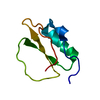 3rdyS S: Starting model for refinement |
|---|---|
| Similar structure data |
-Links
 PDBj
PDBj- Assembly
| Deposited unit | 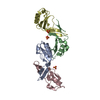
| ||||||||
|---|---|---|---|---|---|---|---|---|---|
| 1 | 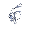
| ||||||||
| 2 | 
| ||||||||
| 3 | 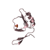
| ||||||||
| 4 | 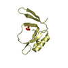
| ||||||||
| Unit cell |
|
-Components
| #1: Protein | Mass: 10169.449 Da / Num. of mol.: 4 Source method: isolated from a genetically manipulated source Source: (gene. exp.) Daucus carota subsp. sativus (plant) / Gene: DCAR_011990Production host:  References: UniProt: A0A166C3G6 #2: Chemical |   Mass: 96.063 Da / Num. of mol.: 3 / Source method: obtained synthetically / Formula: SO4 Mass: 96.063 Da / Num. of mol.: 3 / Source method: obtained synthetically / Formula: SO4#3: Water | ChemComp-HOH / |  Mass: 18.015 Da / Num. of mol.: 42 / Source method: isolated from a natural source / Formula: H2O Mass: 18.015 Da / Num. of mol.: 42 / Source method: isolated from a natural source / Formula: H2OHas ligand of interest | N | Has protein modification | Y | |
|---|
-Experimental details
-Experiment
| Experiment | Method: X-RAY DIFFRACTION / Number of used crystals: 1 |
|---|
- Sample preparation
| Crystal | Density Matthews: 2.66 Å3/Da / Density % sol: 53.84 % |
|---|---|
| Crystal grow | Temperature: 289.15 K / Method: vapor diffusion, sitting drop Details: 0.2 M Sodium sulfate decahydrate, 20% (w/v) Polyethylene glycol (PEG) 3350 |
-Data collection
| Diffraction | Mean temperature: 100 K / Serial crystal experiment: N |
|---|---|
| Diffraction source | Source: SYNCHROTRON / Site: SSRF / Beamline: BL17U1 / Wavelength: 0.97918 Å |
| Detector | Type: DECTRIS EIGER X 16M / Detector: PIXEL / Date: Nov 1, 2020 |
| Radiation | Protocol: SINGLE WAVELENGTH / Monochromatic (M) / Laue (L): M / Scattering type: x-ray |
| Radiation wavelength | Wavelength: 0.97918 Å / Relative weight: 1 |
| Reflection | Resolution: 2.56→44.78 Å / Num. obs: 14451 / % possible obs: 99.1 % / Redundancy: 9.5 % / CC1/2: 0.975 / Rmerge(I) obs: 0.2 / Net I/σ(I): 14.8 |
| Reflection shell | Resolution: 2.56→2.63 Å / Rmerge(I) obs: 0.605 / Mean I/σ(I) obs: 2.4 / Num. unique obs: 955 / CC1/2: 0.597 / % possible all: 93.5 |
- Processing
| Software |
| |||||||||||||||||||||||||||||||||||||||||||||||||||||||||||||||||||||||||||||||||||||||||||||||||||||||||||||||||||||||||||||
|---|---|---|---|---|---|---|---|---|---|---|---|---|---|---|---|---|---|---|---|---|---|---|---|---|---|---|---|---|---|---|---|---|---|---|---|---|---|---|---|---|---|---|---|---|---|---|---|---|---|---|---|---|---|---|---|---|---|---|---|---|---|---|---|---|---|---|---|---|---|---|---|---|---|---|---|---|---|---|---|---|---|---|---|---|---|---|---|---|---|---|---|---|---|---|---|---|---|---|---|---|---|---|---|---|---|---|---|---|---|---|---|---|---|---|---|---|---|---|---|---|---|---|---|---|---|---|
| Refinement | Method to determine structure: MOLECULAR REPLACEMENT Starting model: 3rdy Resolution: 2.56→44.78 Å / SU ML: 0.35 / Cross valid method: THROUGHOUT / σ(F): 1.34 / Phase error: 28.49 / Stereochemistry target values: ML
| |||||||||||||||||||||||||||||||||||||||||||||||||||||||||||||||||||||||||||||||||||||||||||||||||||||||||||||||||||||||||||||
| Solvent computation | Shrinkage radii: 0.9 Å / VDW probe radii: 1.11 Å / Solvent model: FLAT BULK SOLVENT MODEL | |||||||||||||||||||||||||||||||||||||||||||||||||||||||||||||||||||||||||||||||||||||||||||||||||||||||||||||||||||||||||||||
| Displacement parameters | Biso max: 108.22 Å2 / Biso mean: 60.2293 Å2 / Biso min: 34.9 Å2 | |||||||||||||||||||||||||||||||||||||||||||||||||||||||||||||||||||||||||||||||||||||||||||||||||||||||||||||||||||||||||||||
| Refinement step | Cycle: final / Resolution: 2.56→44.78 Å
| |||||||||||||||||||||||||||||||||||||||||||||||||||||||||||||||||||||||||||||||||||||||||||||||||||||||||||||||||||||||||||||
| LS refinement shell | Refine-ID: X-RAY DIFFRACTION / Rfactor Rfree error: 0 / Total num. of bins used: 10
| |||||||||||||||||||||||||||||||||||||||||||||||||||||||||||||||||||||||||||||||||||||||||||||||||||||||||||||||||||||||||||||
| Refinement TLS params. | Method: refined / Refine-ID: X-RAY DIFFRACTION
| |||||||||||||||||||||||||||||||||||||||||||||||||||||||||||||||||||||||||||||||||||||||||||||||||||||||||||||||||||||||||||||
| Refinement TLS group |
|
