 Movie
Movie Controller

[English] 日本語
 Yorodumi
- PDB-7xgq: Crystal structure of chitosanase crystallized by ammonium sulfate... -
+ Open data

- Basic information
| Entry | Database: PDB / ID: 7xgq | ||||||
|---|---|---|---|---|---|---|---|
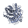
| Title | Crystal structure of chitosanase crystallized by ammonium sulfate with glycan | ||||||
 Components Components | chitosanase | ||||||
 Keywords Keywords | HYDROLASE / glucanase | ||||||
| Biological species |  | ||||||
| Method |  X-RAY DIFFRACTION / SYNCHROTRON / MOLECULAR REPLACEMENT / Resolution: 1.5 Å X-RAY DIFFRACTION / SYNCHROTRON / MOLECULAR REPLACEMENT / Resolution: 1.5 Å | ||||||
 Authors Authors | Guo, Y. / Hoshino, T. | ||||||
| Funding support |  Japan, 1items Japan, 1items
| ||||||
 Citation Citation | Journal: Cryst.Growth Des. / Year: 2022 Title: Influence of Glycan Agents on Protein Crystallization with Ammonium Sulfate Authors: Guo, Y. / Hoshino, T. | ||||||
| History |
|
- Structure visualization
| Structure viewer | Molecule:  MolmilJmol/JSmol MolmilJmol/JSmol |
|---|
- Downloads & links
-Download
| PDBx/mmCIF format | 7xgq.cif.gz | 174.2 KB | Display | PDBx/mmCIF format |
|---|---|---|---|---|
| PDB format | pdb7xgq.ent.gz | 136.3 KB | Display | PDB format |
| PDBx/mmJSON format | 7xgq.json.gz | Tree view | PDBx/mmJSON format | |
| Others |  Other downloads Other downloads |
-Validation report
| Arichive directory | https://data.pdbj.org/pub/pdb/validation_reports/xg/7xgqftp://data.pdbj.org/pub/pdb/validation_reports/xg/7xgq | HTTPS FTP |
|---|
-Related structure data

-Links
 PDBj
PDBj- Assembly
| Deposited unit | 
| ||||||||
|---|---|---|---|---|---|---|---|---|---|
| 1 | 
| ||||||||
| 2 | 
| ||||||||
| Unit cell |
|
-Components
| #1: Protein | Mass: 44594.703 Da / Num. of mol.: 2 Source method: isolated from a genetically manipulated source Details: Sequence used in this structure is identical with the sequence of chain A from structure 7CJU. Source: (gene. exp.) #2: Water | ChemComp-HOH / |  Mass: 18.015 Da / Num. of mol.: 597 / Source method: isolated from a natural source / Formula: H2O Mass: 18.015 Da / Num. of mol.: 597 / Source method: isolated from a natural source / Formula: H2O |
|---|
-Experimental details
-Experiment
| Experiment | Method: X-RAY DIFFRACTION / Number of used crystals: 1 |
|---|
- Sample preparation
| Crystal | Density Matthews: 2.6 Å3/Da / Density % sol: 52.64 % / Mosaicity: 0.22 ° |
|---|---|
| Crystal grow | Temperature: 291 K / Method: vapor diffusion, sitting drop / pH: 5 Details: 0.1M Sodium Citrate, 3.4M Ammonium Sulfate, 5%(v/v) tri[(O-beta-D-glucopyranosyl)ethyloxy]cholane |
-Data collection
| Diffraction | Mean temperature: 100 K / Serial crystal experiment: N |
|---|---|
| Diffraction source | Source: SYNCHROTRON / Site: Photon Factory / Beamline: BL-17A / Wavelength: 1 Å |
| Detector | Type: DECTRIS EIGER X 16M / Detector: PIXEL / Date: Feb 22, 2020 / Details: mirrors |
| Radiation | Monochromator: Si(111) / Protocol: SINGLE WAVELENGTH / Monochromatic (M) / Laue (L): M / Scattering type: x-ray |
| Radiation wavelength | Wavelength: 1 Å / Relative weight: 1 |
| Reflection | Resolution: 1.5→48.73 Å / Num. obs: 142580 / % possible obs: 95.7 % / Redundancy: 6.8 % / CC1/2: 0.991 / Rmerge(I) obs: 0.056 / Rpim(I) all: 0.023 / Rrim(I) all: 0.061 / Net I/σ(I): 18.8 |
| Reflection shell | Resolution: 1.5→1.53 Å / Redundancy: 6.9 % / Rmerge(I) obs: 0.975 / Num. unique obs: 6809 / CC1/2: 0.559 / Rpim(I) all: 0.397 / Rrim(I) all: 1.055 / % possible all: 93.1 |
- Processing
| Software |
| ||||||||||||||||||||||||
|---|---|---|---|---|---|---|---|---|---|---|---|---|---|---|---|---|---|---|---|---|---|---|---|---|---|
| Refinement | Method to determine structure: MOLECULAR REPLACEMENT Starting model: 7CJU Resolution: 1.5→48.729 Å / SU ML: 0.19 / Cross valid method: THROUGHOUT / σ(F): 1.92 / Phase error: 22.19 / Stereochemistry target values: ML
| ||||||||||||||||||||||||
| Solvent computation | Shrinkage radii: 0.9 Å / VDW probe radii: 1.11 Å / Solvent model: FLAT BULK SOLVENT MODEL | ||||||||||||||||||||||||
| Displacement parameters | Biso max: 69.67 Å2 / Biso mean: 22.4989 Å2 / Biso min: 9.27 Å2 | ||||||||||||||||||||||||
| Refinement step | Cycle: final / Resolution: 1.5→48.729 Å
| ||||||||||||||||||||||||
| LS refinement shell | Resolution: 1.5→1.53 Å
|
