 Movie
Movie Controller


+ Open data

- Basic information
| Entry | Database: PDB / ID: 7xg7 | ||||||
|---|---|---|---|---|---|---|---|
| Title | Crystal structure of PstS protein from cyanophage Syn19 | ||||||
 Components Components | Phosphate transporter subunit | ||||||
 Keywords Keywords | TRANSPORT PROTEIN / phosphate binding protein / cyanobacteria / cyanophage | ||||||
| Function / homology |  Function and homology information Function and homology informationphosphate ion transmembrane transport / phosphate ion binding / ATP-binding cassette (ABC) transporter complex Similarity search - Function | ||||||
| Biological species |  Synechococcus phage Syn19 (virus) Synechococcus phage Syn19 (virus) | ||||||
| Method |  X-RAY DIFFRACTION / SYNCHROTRON / MOLECULAR REPLACEMENT / Resolution: 1.7 Å X-RAY DIFFRACTION / SYNCHROTRON / MOLECULAR REPLACEMENT / Resolution: 1.7 Å | ||||||
 Authors Authors | Cai, K. / Jiang, Y.L. | ||||||
| Funding support | 1items
| ||||||
 Citation Citation | Journal: Environ.Microbiol. / Year: 2022 Title: Biochemical and structural characterization of the cyanophage-encoded phosphate-binding protein: implications for enhanced phosphate uptake of infected cyanobacteria Authors: Zhao, F. / Lin, X. / Cai, K. / Jiang, Y. / Ni, T. / Chen, Y. / Feng, J. / Dang, S. / Zhou, C.Z. / Zeng, Q. | ||||||
| History |
|
- Structure visualization
| Structure viewer | Molecule: MolmilJmol/JSmol |
|---|
- Downloads & links
-Download
| PDBx/mmCIF format | 7xg7.cif.gz | 129.4 KB | Display | PDBx/mmCIF format |
|---|---|---|---|---|
| PDB format | pdb7xg7.ent.gz | 99.9 KB | Display | PDB format |
| PDBx/mmJSON format | 7xg7.json.gz | Tree view | PDBx/mmJSON format | |
| Others |  Other downloads Other downloads |
-Validation report
| Summary document | 7xg7_validation.pdf.gz | 1.2 MB | Display | wwPDB validaton report |
|---|---|---|---|---|
| Full document | 7xg7_full_validation.pdf.gz | 1.2 MB | Display | |
| Data in XML | 7xg7_validation.xml.gz | 26.3 KB | Display | |
| Data in CIF | 7xg7_validation.cif.gz | 38.1 KB | Display | |
| Arichive directory | https://data.pdbj.org/pub/pdb/validation_reports/xg/7xg7ftp://data.pdbj.org/pub/pdb/validation_reports/xg/7xg7 | HTTPS FTP |
-Related structure data
| Related structure data |  7xg8C 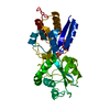 2abhS S: Starting model for refinement C: citing same article ( |
|---|---|
| Similar structure data |
-Links
 PDBj
PDBj


- Assembly
| Deposited unit | 
| ||||||||
|---|---|---|---|---|---|---|---|---|---|
| 1 | 
| ||||||||
| 2 | 
| ||||||||
| Unit cell |
|
-Components
| #1: Protein | Mass: 34119.496 Da / Num. of mol.: 2 Source method: isolated from a genetically manipulated source Source: (gene. exp.) Synechococcus phage Syn19 (virus) / Gene: pstS, Syn19_159 / Production host:  #2: Chemical |   Mass: 94.971 Da / Num. of mol.: 2 / Source method: obtained synthetically / Formula: PO4 / Feature type: SUBJECT OF INVESTIGATION Mass: 94.971 Da / Num. of mol.: 2 / Source method: obtained synthetically / Formula: PO4 / Feature type: SUBJECT OF INVESTIGATION#3: Chemical | ChemComp-GOL /   Mass: 92.094 Da / Num. of mol.: 5 / Source method: obtained synthetically / Formula: C3H8O3 Mass: 92.094 Da / Num. of mol.: 5 / Source method: obtained synthetically / Formula: C3H8O3#4: Water | ChemComp-HOH / |  Mass: 18.015 Da / Num. of mol.: 359 / Source method: isolated from a natural source / Formula: H2O Mass: 18.015 Da / Num. of mol.: 359 / Source method: isolated from a natural source / Formula: H2OHas ligand of interest | Y | Has protein modification | Y | |
|---|
-Experimental details
-Experiment
| Experiment | Method: X-RAY DIFFRACTION / Number of used crystals: 1 |
|---|
- Sample preparation
| Crystal | Density Matthews: 2.68 Å3/Da / Density % sol: 54.15 % |
|---|---|
| Crystal grow | Temperature: 289 K / Method: vapor diffusion, hanging drop Details: 18% (w/v) polyethylene glycol 3350, 0.2 M sodium formate |
-Data collection
| Diffraction | Mean temperature: 100 K / Serial crystal experiment: N |
|---|---|
| Diffraction source | Source: SYNCHROTRON / Site: SSRF  / Beamline: BL17U / Wavelength: 0.97918 Å / Beamline: BL17U / Wavelength: 0.97918 Å |
| Detector | Type: DECTRIS EIGER X 16M / Detector: PIXEL / Date: Mar 23, 2016 |
| Radiation | Protocol: SINGLE WAVELENGTH / Monochromatic (M) / Laue (L): M / Scattering type: x-ray |
| Radiation wavelength | Wavelength: 0.97918 Å / Relative weight: 1 |
| Reflection | Resolution: 1.7→50 Å / Num. obs: 79022 / % possible obs: 98.8 % / Redundancy: 3.1 % / Rmerge(I) obs: 0.064 / Rsym value: 0.064 / Net I/σ(I): 14.247 |
| Reflection shell | Resolution: 1.7→1.76 Å / Rmerge(I) obs: 0.413 / Mean I/σ(I) obs: 2.658 / Num. unique obs: 7809 / Rsym value: 0.413 / % possible all: 98.5 |
- Processing
| Software |
| ||||||||||||||||||||||||||||||||||||||||||||||||||||||||||||
|---|---|---|---|---|---|---|---|---|---|---|---|---|---|---|---|---|---|---|---|---|---|---|---|---|---|---|---|---|---|---|---|---|---|---|---|---|---|---|---|---|---|---|---|---|---|---|---|---|---|---|---|---|---|---|---|---|---|---|---|---|---|
| Refinement | Method to determine structure: MOLECULAR REPLACEMENT Starting model: 2abh Resolution: 1.7→50 Å / Cor.coef. Fo:Fc: 0.969 / Cor.coef. Fo:Fc free: 0.964 / SU B: 1.829 / SU ML: 0.059 / Cross valid method: THROUGHOUT / σ(F): 0 / ESU R: 0.084 / ESU R Free: 0.08 / Stereochemistry target values: MAXIMUM LIKELIHOOD Details: HYDROGENS HAVE BEEN ADDED IN THE RIDING POSITIONS U VALUES : REFINED INDIVIDUALLY
| ||||||||||||||||||||||||||||||||||||||||||||||||||||||||||||
| Solvent computation | Ion probe radii: 0.8 Å / Shrinkage radii: 0.8 Å / VDW probe radii: 1.2 Å / Solvent model: MASK | ||||||||||||||||||||||||||||||||||||||||||||||||||||||||||||
| Displacement parameters | Biso max: 137.94 Å2 / Biso mean: 21.122 Å2 / Biso min: 6.3 Å2
| ||||||||||||||||||||||||||||||||||||||||||||||||||||||||||||
| Refinement step | Cycle: final / Resolution: 1.7→50 Å
| ||||||||||||||||||||||||||||||||||||||||||||||||||||||||||||
| Refine LS restraints |
| ||||||||||||||||||||||||||||||||||||||||||||||||||||||||||||
| LS refinement shell | Resolution: 1.7→1.743 Å / Rfactor Rfree error: 0
|
