 Movie
Movie Controller

[English] 日本語
 Yorodumi
- PDB-7xfr: Crystal structure of WIPI2b in complex with the second site of ATG16L1 -
+ Open data

- Basic information
| Entry | Database: PDB / ID: 7xfr | ||||||
|---|---|---|---|---|---|---|---|
| Title | Crystal structure of WIPI2b in complex with the second site of ATG16L1 | ||||||
 Components Components |
| ||||||
 Keywords Keywords | PROTEIN BINDING / WIPI2b / ATG16L1 | ||||||
| Function / homology |  Function and homology information Function and homology informationC-terminal protein lipidation / Atg12-Atg5-Atg16 complex / negative regulation of dendrite extension / vacuole-isolation membrane contact site / ubiquitin-like protein transferase activity / microautophagy / dendrite arborization / xenophagy / corpus callosum development / protein localization to phagophore assembly site ...C-terminal protein lipidation / Atg12-Atg5-Atg16 complex / negative regulation of dendrite extension / vacuole-isolation membrane contact site / ubiquitin-like protein transferase activity / microautophagy / dendrite arborization / xenophagy / corpus callosum development / protein localization to phagophore assembly site / phagophore assembly site membrane / negative stranded viral RNA replication / endolysosome membrane / axonal transport / Macroautophagy / autophagosome membrane / axoneme / autophagosome assembly / protein-membrane adaptor activity / positive regulation of autophagy / autophagosome / hippocampus development / macroautophagy / protein transport / GTPase binding / sperm midpiece / defense response to virus / axon / glutamatergic synapse / identical protein binding / cytosol Similarity search - Function | ||||||
| Biological species |  Homo sapiens (human) Homo sapiens (human) | ||||||
| Method |  X-RAY DIFFRACTION / SYNCHROTRON / MOLECULAR REPLACEMENT / Resolution: 1.76 Å X-RAY DIFFRACTION / SYNCHROTRON / MOLECULAR REPLACEMENT / Resolution: 1.76 Å | ||||||
 Authors Authors | Gong, X.Y. / Pan, L.F. | ||||||
| Funding support |  China, 1items China, 1items
| ||||||
 Citation Citation | Journal: Sci Adv / Year: 2023 Title: ATG16L1 adopts a dual-binding site mode to interact with WIPI2b in autophagy. Authors: Gong, X. / Wang, Y. / Tang, Y. / Wang, Y. / Zhang, M. / Li, M. / Zhang, Y. / Pan, L. | ||||||
| History |
|
- Structure visualization
| Structure viewer | Molecule: MolmilJmol/JSmol |
|---|
- Downloads & links
-Download
| PDBx/mmCIF format | 7xfr.cif.gz | 462.5 KB | Display | PDBx/mmCIF format |
|---|---|---|---|---|
| PDB format | pdb7xfr.ent.gz | 370.2 KB | Display | PDB format |
| PDBx/mmJSON format | 7xfr.json.gz | Tree view | PDBx/mmJSON format | |
| Others |  Other downloads Other downloads |
-Validation report
| Arichive directory | https://data.pdbj.org/pub/pdb/validation_reports/xf/7xfrftp://data.pdbj.org/pub/pdb/validation_reports/xf/7xfr | HTTPS FTP |
|---|
-Related structure data
| Related structure data |  7f69C  5ltgS S: Starting model for refinement C: citing same article ( |
|---|---|
| Similar structure data |
-Links
 PDBj
PDBj- Assembly
| Deposited unit | 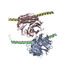
| ||||||||||||
|---|---|---|---|---|---|---|---|---|---|---|---|---|---|
| 1 | 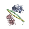
| ||||||||||||
| Unit cell |
|
-Components
| #1: Protein | Mass: 35310.637 Da / Num. of mol.: 2 Source method: isolated from a genetically manipulated source Source: (gene. exp.) Homo sapiens (human) / Gene: WIPI2, CGI-50 / Production host:  #2: Protein | Mass: 7815.774 Da / Num. of mol.: 2 Source method: isolated from a genetically manipulated source Source: (gene. exp.) Homo sapiens (human) / Gene: ATG16L1, APG16L, UNQ9393/PRO34307 / Production host: #3: Water | ChemComp-HOH / |  Mass: 18.015 Da / Num. of mol.: 424 / Source method: isolated from a natural source / Formula: H2O Mass: 18.015 Da / Num. of mol.: 424 / Source method: isolated from a natural source / Formula: H2O |
|---|
-Experimental details
-Experiment
| Experiment | Method: X-RAY DIFFRACTION / Number of used crystals: 1 |
|---|
- Sample preparation
| Crystal | Density Matthews: 2.68 Å3/Da / Density % sol: 54.03 % |
|---|---|
| Crystal grow | Temperature: 289 K / Method: vapor diffusion, sitting drop / pH: 6.5 Details: 0.1 M MES monohydrate(pH 6.5), 12% w/v Polyethylene glycol 20000 |
-Data collection
| Diffraction | Mean temperature: 100 K / Serial crystal experiment: N |
|---|---|
| Diffraction source | Source: SYNCHROTRON / Site: SSRF / Beamline: BL17U1 / Wavelength: 0.97918 Å |
| Detector | Type: ADSC QUANTUM 315r / Detector: CCD / Date: Nov 1, 2020 |
| Radiation | Protocol: SINGLE WAVELENGTH / Monochromatic (M) / Laue (L): M / Scattering type: x-ray |
| Radiation wavelength | Wavelength: 0.97918 Å / Relative weight: 1 |
| Reflection | Resolution: 1.76→73.59 Å / Num. obs: 83257 / % possible obs: 93.71 % / Redundancy: 6.7 % / Biso Wilson estimate: 31.73 Å2 / Rrim(I) all: 0.037 / Net I/av σ(I): 24.4 / Net I/σ(I): 24.71 |
| Reflection shell | Resolution: 1.76→1.79 Å / Num. unique obs: 4440 / Rrim(I) all: 0.748 |
- Processing
| Software |
| ||||||||||||||||||||||||||||||||||||||||||||||||||||||||||||||||||||||||||||||||||||||||||||||||||||||||||||||||||||||||||||||||||||||||||||||||||||||||||||||||||||||||||||||||||||||||||||||||||||||||||||||||||
|---|---|---|---|---|---|---|---|---|---|---|---|---|---|---|---|---|---|---|---|---|---|---|---|---|---|---|---|---|---|---|---|---|---|---|---|---|---|---|---|---|---|---|---|---|---|---|---|---|---|---|---|---|---|---|---|---|---|---|---|---|---|---|---|---|---|---|---|---|---|---|---|---|---|---|---|---|---|---|---|---|---|---|---|---|---|---|---|---|---|---|---|---|---|---|---|---|---|---|---|---|---|---|---|---|---|---|---|---|---|---|---|---|---|---|---|---|---|---|---|---|---|---|---|---|---|---|---|---|---|---|---|---|---|---|---|---|---|---|---|---|---|---|---|---|---|---|---|---|---|---|---|---|---|---|---|---|---|---|---|---|---|---|---|---|---|---|---|---|---|---|---|---|---|---|---|---|---|---|---|---|---|---|---|---|---|---|---|---|---|---|---|---|---|---|---|---|---|---|---|---|---|---|---|---|---|---|---|---|---|---|---|
| Refinement | Method to determine structure: MOLECULAR REPLACEMENT Starting model: 5LTG Resolution: 1.76→24.99 Å / SU ML: 0.1383 / Cross valid method: FREE R-VALUE / σ(F): 1.35 / Phase error: 18.7137 Stereochemistry target values: GeoStd + Monomer Library + CDL v1.2
| ||||||||||||||||||||||||||||||||||||||||||||||||||||||||||||||||||||||||||||||||||||||||||||||||||||||||||||||||||||||||||||||||||||||||||||||||||||||||||||||||||||||||||||||||||||||||||||||||||||||||||||||||||
| Solvent computation | Shrinkage radii: 0.9 Å / VDW probe radii: 1.11 Å / Solvent model: FLAT BULK SOLVENT MODEL | ||||||||||||||||||||||||||||||||||||||||||||||||||||||||||||||||||||||||||||||||||||||||||||||||||||||||||||||||||||||||||||||||||||||||||||||||||||||||||||||||||||||||||||||||||||||||||||||||||||||||||||||||||
| Displacement parameters | Biso mean: 45.27 Å2 | ||||||||||||||||||||||||||||||||||||||||||||||||||||||||||||||||||||||||||||||||||||||||||||||||||||||||||||||||||||||||||||||||||||||||||||||||||||||||||||||||||||||||||||||||||||||||||||||||||||||||||||||||||
| Refinement step | Cycle: LAST / Resolution: 1.76→24.99 Å
| ||||||||||||||||||||||||||||||||||||||||||||||||||||||||||||||||||||||||||||||||||||||||||||||||||||||||||||||||||||||||||||||||||||||||||||||||||||||||||||||||||||||||||||||||||||||||||||||||||||||||||||||||||
| Refine LS restraints |
| ||||||||||||||||||||||||||||||||||||||||||||||||||||||||||||||||||||||||||||||||||||||||||||||||||||||||||||||||||||||||||||||||||||||||||||||||||||||||||||||||||||||||||||||||||||||||||||||||||||||||||||||||||
| LS refinement shell |
|
