 Movie
Movie Controller


+ Open data

- Basic information
| Entry | Database: PDB / ID: 7xdh | |||||||||||||||||||||||||||||||
|---|---|---|---|---|---|---|---|---|---|---|---|---|---|---|---|---|---|---|---|---|---|---|---|---|---|---|---|---|---|---|---|---|
| Title | Crystal structure of a dimeric interlocked parallel G-quadruplex | |||||||||||||||||||||||||||||||
 Components Components | DNA (5'-D(* Keywords KeywordsDNA / G-quadruplex / interlocked | Function / homology | : / DNA / DNA (> 10) |  Function and homology information Function and homology informationBiological species | synthetic construct (others) | Method |  X-RAY DIFFRACTION / SYNCHROTRON / MOLECULAR REPLACEMENT / Resolution: 1.83 Å X-RAY DIFFRACTION / SYNCHROTRON / MOLECULAR REPLACEMENT / Resolution: 1.83 Å  Authors AuthorsNgo, K.H. / Liew, C.W. / Lattmann, S. / Winnerdy, F.R. / Phan, A.T. | Funding support | |  Singapore, 2items Singapore, 2items
 CitationJournal: Biochem.Biophys.Res.Commun. / Year: 2022 CitationJournal: Biochem.Biophys.Res.Commun. / Year: 2022Title: Crystal structures of an HIV-1 integrase aptamer: Formation of a water-mediated A.G.G.G.G pentad in an interlocked G-quadruplex. Authors: Ngo, K.H. / Liew, C.W. / Lattmann, S. / Winnerdy, F.R. / Phan, A.T. History |
|
- Structure visualization
| Structure viewer | Molecule: MolmilJmol/JSmol |
|---|
- Downloads & links
-Download
| PDBx/mmCIF format | 7xdh.cif.gz | 98.1 KB | Display | PDBx/mmCIF format |
|---|---|---|---|---|
| PDB format | pdb7xdh.ent.gz | 63.8 KB | Display | PDB format |
| PDBx/mmJSON format | 7xdh.json.gz | Tree view | PDBx/mmJSON format | |
| Others |  Other downloads Other downloads |
-Validation report
| Arichive directory | https://data.pdbj.org/pub/pdb/validation_reports/xd/7xdhftp://data.pdbj.org/pub/pdb/validation_reports/xd/7xdh | HTTPS FTP |
|---|
-Related structure data
| Related structure data |  7x7gC  7xh9C  7xhdC  7xieC 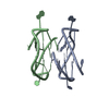 1y8dS S: Starting model for refinement C: citing same article ( |
|---|---|
| Similar structure data |
-Links
 PDBj
PDBj


- Assembly
| Deposited unit | 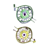
| ||||||||||||
|---|---|---|---|---|---|---|---|---|---|---|---|---|---|
| 1 | 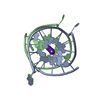
| ||||||||||||
| 2 | 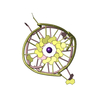
| ||||||||||||
| Unit cell |
|
-Components
| #1: DNA chain | Mass: 5140.312 Da / Num. of mol.: 4 / Source method: obtained synthetically / Source: (synth.) synthetic construct (others) #2: Chemical | ChemComp-K /   Mass: 39.098 Da / Num. of mol.: 10 / Source method: obtained synthetically / Formula: K / Feature type: SUBJECT OF INVESTIGATION Mass: 39.098 Da / Num. of mol.: 10 / Source method: obtained synthetically / Formula: K / Feature type: SUBJECT OF INVESTIGATION#3: Water | ChemComp-HOH / |  Mass: 18.015 Da / Num. of mol.: 49 / Source method: isolated from a natural source / Formula: H2O Mass: 18.015 Da / Num. of mol.: 49 / Source method: isolated from a natural source / Formula: H2OHas ligand of interest | Y | |
|---|
-Experimental details
-Experiment
| Experiment | Method: X-RAY DIFFRACTION / Number of used crystals: 1 |
|---|
- Sample preparation
| Crystal | Density Matthews: 2.05 Å3/Da / Density % sol: 40.03 % |
|---|---|
| Crystal grow | Temperature: 293 K / Method: vapor diffusion, sitting drop Details: 0.08 M Sodium chloride, 0.02 M Magnesium chloride hexahydrate, 0.04 M Sodium cacodylate trihydrate pH 7.0, 40% v/v (+/-)-2-Methyl-2,4-pentanediol, 0.012 M Spermine tetrahydrochloride |
-Data collection
| Diffraction | Mean temperature: 90 K / Serial crystal experiment: N |
|---|---|
| Diffraction source | Source: SYNCHROTRON / Site: Australian Synchrotron  / Beamline: MX2 / Wavelength: 0.953659 Å / Beamline: MX2 / Wavelength: 0.953659 Å |
| Detector | Type: DECTRIS EIGER X 9M / Detector: PIXEL / Date: Mar 19, 2022 |
| Radiation | Protocol: SINGLE WAVELENGTH / Monochromatic (M) / Laue (L): M / Scattering type: x-ray |
| Radiation wavelength | Wavelength: 0.953659 Å / Relative weight: 1 |
| Reflection | Resolution: 1.826→29.26 Å / Num. obs: 13916 / % possible obs: 95.01 % / Redundancy: 4.2 % / Biso Wilson estimate: 35.69 Å2 / CC1/2: 0.999 / Net I/σ(I): 11.98 |
| Reflection shell | Resolution: 1.826→1.857 Å / Num. unique obs: 1413 / CC1/2: 0.962 |
- Processing
| Software |
| |||||||||||||||||||||||||||||||||||||||||||||||||||||||||||||||||||||||||||||
|---|---|---|---|---|---|---|---|---|---|---|---|---|---|---|---|---|---|---|---|---|---|---|---|---|---|---|---|---|---|---|---|---|---|---|---|---|---|---|---|---|---|---|---|---|---|---|---|---|---|---|---|---|---|---|---|---|---|---|---|---|---|---|---|---|---|---|---|---|---|---|---|---|---|---|---|---|---|---|
| Refinement | Method to determine structure: MOLECULAR REPLACEMENT Starting model: 1Y8D Resolution: 1.83→29.26 Å / SU ML: 0.2319 / Cross valid method: FREE R-VALUE / σ(F): 2 / Phase error: 37.2734 Stereochemistry target values: GeoStd + Monomer Library + CDL v1.2
| |||||||||||||||||||||||||||||||||||||||||||||||||||||||||||||||||||||||||||||
| Solvent computation | Shrinkage radii: 0.9 Å / VDW probe radii: 1.11 Å / Solvent model: FLAT BULK SOLVENT MODEL | |||||||||||||||||||||||||||||||||||||||||||||||||||||||||||||||||||||||||||||
| Displacement parameters | Biso mean: 50.94 Å2 | |||||||||||||||||||||||||||||||||||||||||||||||||||||||||||||||||||||||||||||
| Refinement step | Cycle: LAST / Resolution: 1.83→29.26 Å
| |||||||||||||||||||||||||||||||||||||||||||||||||||||||||||||||||||||||||||||
| Refine LS restraints |
| |||||||||||||||||||||||||||||||||||||||||||||||||||||||||||||||||||||||||||||
| LS refinement shell |
| |||||||||||||||||||||||||||||||||||||||||||||||||||||||||||||||||||||||||||||
| Refinement TLS params. | Method: refined / Origin x: 4.84774730784 Å / Origin y: -2.51563985782 Å / Origin z: -24.786644211 Å
| |||||||||||||||||||||||||||||||||||||||||||||||||||||||||||||||||||||||||||||
| Refinement TLS group | Selection details: all |
