 Movie
Movie Controller


+ Open data

- Basic information
| Entry | Database: PDB / ID: 7xcm | ||||||
|---|---|---|---|---|---|---|---|
| Title | Crystal structure of sulfite MttB structure at 3.2 A resolution | ||||||
 Components Components | Trimethylamine methyltransferase | ||||||
 Keywords Keywords | TRANSFERASE / pyrrolysine / trimethylamine / methyltransferase / corrinoid protein | ||||||
| Function / homology |  Function and homology information Function and homology informationtrimethylamine-corrinoid protein Co-methyltransferase / trimethylamine methyltransferase activity / methanogenesis / methylation Similarity search - Function | ||||||
| Biological species |  Methanosarcina barkeri MS (archaea) Methanosarcina barkeri MS (archaea) | ||||||
| Method |  X-RAY DIFFRACTION / MOLECULAR REPLACEMENT / Resolution: 3.2 Å X-RAY DIFFRACTION / MOLECULAR REPLACEMENT / Resolution: 3.2 Å | ||||||
 Authors Authors | Li, J. / Chan, M.K. | ||||||
| Funding support |  Hong Kong, 1items Hong Kong, 1items
| ||||||
 Citation Citation | Journal: Commun Biol / Year: 2023 Title: Insights into pyrrolysine function from structures of a trimethylamine methyltransferase and its corrinoid protein complex. Authors: Li, J. / Kang, P.T. / Jiang, R. / Lee, J.Y. / Soares, J.A. / Krzycki, J.A. / Chan, M.K. | ||||||
| History |
|
- Structure visualization
| Structure viewer | Molecule: MolmilJmol/JSmol |
|---|
- Downloads & links
-Download
| PDBx/mmCIF format | 7xcm.cif.gz | 561.3 KB | Display | PDBx/mmCIF format |
|---|---|---|---|---|
| PDB format | pdb7xcm.ent.gz | 463.6 KB | Display | PDB format |
| PDBx/mmJSON format | 7xcm.json.gz | Tree view | PDBx/mmJSON format | |
| Others |  Other downloads Other downloads |
-Validation report
| Arichive directory | https://data.pdbj.org/pub/pdb/validation_reports/xc/7xcmftp://data.pdbj.org/pub/pdb/validation_reports/xc/7xcm | HTTPS FTP |
|---|
-Related structure data
| Related structure data | 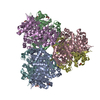 7xclSC  7xcnC S: Starting model for refinement C: citing same article ( |
|---|---|
| Similar structure data |
-Links
 PDBj
PDBj- Assembly
| Deposited unit | 
| ||||||||
|---|---|---|---|---|---|---|---|---|---|
| 1 |
| ||||||||
| Unit cell |
|
-Components
| #1: Protein | Mass: 54746.387 Da / Num. of mol.: 6 Source method: isolated from a genetically manipulated source Source: (gene. exp.) Methanosarcina barkeri MS (archaea) / Gene: MSBRM_0461 / Production host: Methanosarcina acetivorans C2A (archaea)References: UniProt: A0A0E3QRM4, trimethylamine-corrinoid protein Co-methyltransferase #2: Chemical | ChemComp-BG3 /   Mass: 209.220 Da / Num. of mol.: 6 / Source method: obtained synthetically / Formula: C6H11NO5S / Feature type: SUBJECT OF INVESTIGATION Mass: 209.220 Da / Num. of mol.: 6 / Source method: obtained synthetically / Formula: C6H11NO5S / Feature type: SUBJECT OF INVESTIGATION#3: Chemical | ChemComp-NA /   Mass: 22.990 Da / Num. of mol.: 6 / Source method: obtained synthetically / Formula: Na Mass: 22.990 Da / Num. of mol.: 6 / Source method: obtained synthetically / Formula: Na#4: Chemical | ChemComp-GOL /   Mass: 92.094 Da / Num. of mol.: 11 / Source method: obtained synthetically / Formula: C3H8O3 Mass: 92.094 Da / Num. of mol.: 11 / Source method: obtained synthetically / Formula: C3H8O3#5: Water | ChemComp-HOH / |  Mass: 18.015 Da / Num. of mol.: 165 / Source method: isolated from a natural source / Formula: H2O Mass: 18.015 Da / Num. of mol.: 165 / Source method: isolated from a natural source / Formula: H2OHas ligand of interest | Y | Has protein modification | Y | |
|---|
-Experimental details
-Experiment
| Experiment | Method: X-RAY DIFFRACTION / Number of used crystals: 1 |
|---|
- Sample preparation
| Crystal | Density Matthews: 4.11 Å3/Da / Density % sol: 70.1 % |
|---|---|
| Crystal grow | Temperature: 283 K / Method: vapor diffusion, hanging drop Details: 1 ul protein (8 mg/mL MttB in 20 mM potassium phosphate buffer pH 7.4, 500 mM NaCl, 240 mM imidazole, 20% glycerol), 1 ul reservoir (4% MPD, 0.1 M citric acid pH 4.5) solutions, reservoir ...Details: 1 ul protein (8 mg/mL MttB in 20 mM potassium phosphate buffer pH 7.4, 500 mM NaCl, 240 mM imidazole, 20% glycerol), 1 ul reservoir (4% MPD, 0.1 M citric acid pH 4.5) solutions, reservoir solution containing saturated sodium dithionite |
-Data collection
| Diffraction | Mean temperature: 100 K / Serial crystal experiment: N |
|---|---|
| Diffraction source | Source: ROTATING ANODE / Type: RIGAKU FR-E+ / Wavelength: 1.54187 Å |
| Detector | Type: RIGAKU RAXIS IV / Detector: IMAGE PLATE / Date: Dec 15, 2018 |
| Radiation | Protocol: SINGLE WAVELENGTH / Monochromatic (M) / Laue (L): M / Scattering type: x-ray |
| Radiation wavelength | Wavelength: 1.54187 Å / Relative weight: 1 |
| Reflection | Resolution: 3.2→20 Å / Num. obs: 74099 / % possible obs: 85.4 % / Redundancy: 2.7 % / Rmerge(I) obs: 0.196 / Net I/σ(I): 2.6 |
| Reflection shell | Resolution: 3.2→3.27 Å / Num. unique obs: 11486 / Rpim(I) all: 0.254 |
- Processing
| Software |
| |||||||||||||||||||||||||||||||||||||||||||||||||||||||||||||||||||||||||||||
|---|---|---|---|---|---|---|---|---|---|---|---|---|---|---|---|---|---|---|---|---|---|---|---|---|---|---|---|---|---|---|---|---|---|---|---|---|---|---|---|---|---|---|---|---|---|---|---|---|---|---|---|---|---|---|---|---|---|---|---|---|---|---|---|---|---|---|---|---|---|---|---|---|---|---|---|---|---|---|
| Refinement | Method to determine structure: MOLECULAR REPLACEMENT Starting model: 7xcl Resolution: 3.2→20 Å / Cross valid method: THROUGHOUT / σ(F): 0
| |||||||||||||||||||||||||||||||||||||||||||||||||||||||||||||||||||||||||||||
| Solvent computation | Bsol: 46.955 Å2 | |||||||||||||||||||||||||||||||||||||||||||||||||||||||||||||||||||||||||||||
| Displacement parameters | Biso max: 149.96 Å2 / Biso mean: 53.0752 Å2 / Biso min: 1 Å2
| |||||||||||||||||||||||||||||||||||||||||||||||||||||||||||||||||||||||||||||
| Refinement step | Cycle: final / Resolution: 3.2→20 Å
| |||||||||||||||||||||||||||||||||||||||||||||||||||||||||||||||||||||||||||||
| Refine LS restraints |
| |||||||||||||||||||||||||||||||||||||||||||||||||||||||||||||||||||||||||||||
| LS refinement shell | Refine-ID: X-RAY DIFFRACTION / Rfactor Rfree error: 0 / Total num. of bins used: 10
| |||||||||||||||||||||||||||||||||||||||||||||||||||||||||||||||||||||||||||||
| Xplor file |
|
