 Movie
Movie Controller

[English] 日本語
 Yorodumi
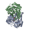
- PDB-7x5n: Crystal structure of E. faecium SHMT in complex with (+)-SHIN-1 a... -
+ Open data

- Basic information
| Entry | Database: PDB / ID: 7x5n | ||||||
|---|---|---|---|---|---|---|---|
| Title | Crystal structure of E. faecium SHMT in complex with (+)-SHIN-1 and PLP-Ser | ||||||
 Components Components | Serine hydroxymethyltransferase | ||||||
 Keywords Keywords | TRANSFERASE / E. faecium / Serine hydroxymethyltransferase / 1C metabolism | ||||||
| Function / homology |  Function and homology information Function and homology informationglycine hydroxymethyltransferase / glycine hydroxymethyltransferase activity / : / tetrahydrofolate interconversion / methyltransferase activity / pyridoxal phosphate binding / methylation / cytosol Similarity search - Function | ||||||
| Biological species |  Enterococcus faecium (bacteria) Enterococcus faecium (bacteria) | ||||||
| Method |  X-RAY DIFFRACTION / SYNCHROTRON / FOURIER SYNTHESIS / Resolution: 1.9 Å X-RAY DIFFRACTION / SYNCHROTRON / FOURIER SYNTHESIS / Resolution: 1.9 Å | ||||||
 Authors Authors | Hasegawa, K. / Hayashi, H. | ||||||
| Funding support | 1items
| ||||||
 Citation Citation | Journal: Commun Biol / Year: 2022 Title: Serine hydroxymethyltransferase as a potential target of antibacterial agents acting synergistically with one-carbon metabolism-related inhibitors. Authors: Makino, Y. / Oe, C. / Iwama, K. / Suzuki, S. / Nishiyama, A. / Hasegawa, K. / Okuda, H. / Hirata, K. / Ueno, M. / Kawaji, K. / Sasano, M. / Usui, E. / Hosaka, T. / Yabuki, Y. / Shirouzu, M. ...Authors: Makino, Y. / Oe, C. / Iwama, K. / Suzuki, S. / Nishiyama, A. / Hasegawa, K. / Okuda, H. / Hirata, K. / Ueno, M. / Kawaji, K. / Sasano, M. / Usui, E. / Hosaka, T. / Yabuki, Y. / Shirouzu, M. / Katsumi, M. / Murayama, K. / Hayashi, H. / Kodama, E.N. | ||||||
| History |
|
- Structure visualization
| Structure viewer | Molecule: MolmilJmol/JSmol |
|---|
- Downloads & links
-Download
| PDBx/mmCIF format | 7x5n.cif.gz | 221.2 KB | Display | PDBx/mmCIF format |
|---|---|---|---|---|
| PDB format | pdb7x5n.ent.gz | 141.9 KB | Display | PDB format |
| PDBx/mmJSON format | 7x5n.json.gz | Tree view | PDBx/mmJSON format | |
| Others |  Other downloads Other downloads |
-Validation report
| Arichive directory | https://data.pdbj.org/pub/pdb/validation_reports/x5/7x5nftp://data.pdbj.org/pub/pdb/validation_reports/x5/7x5n | HTTPS FTP |
|---|
-Related structure data

-Links
 PDBj
PDBj- Assembly
| Deposited unit | 
| ||||||||||||
|---|---|---|---|---|---|---|---|---|---|---|---|---|---|
| 1 |
| ||||||||||||
| Unit cell |
|
-Components
| #1: Protein | Mass: 45005.035 Da / Num. of mol.: 2 Source method: isolated from a genetically manipulated source Source: (gene. exp.) Enterococcus faecium (bacteria)Gene: glyA, glyA_1, B1P95_02920, BU183_09275, BU187_13395, BU190_13075, BU192_03790, BXT96_08900, CQR37_06830, CUM68_02790, CUN04_11625, CUS10_03200, CWC53_10155, DKP91_07450, DTPHA_1400422, DTPHA_ ...Gene: glyA, glyA_1, B1P95_02920, BU183_09275, BU187_13395, BU190_13075, BU192_03790, BXT96_08900, CQR37_06830, CUM68_02790, CUN04_11625, CUS10_03200, CWC53_10155, DKP91_07450, DTPHA_1400422, DTPHA_600996, EB12_01905, EfmAA708_21800, F6440_11360, GBM44_11330, GBM73_12625, GJ652_13105, SAMEA3893517_00378 Production host: References: UniProt: A0A133CK16, glycine hydroxymethyltransferase #2: Chemical | 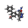  Mass: 400.473 Da / Num. of mol.: 2 / Source method: obtained synthetically / Formula: C24H24N4O2 / Feature type: SUBJECT OF INVESTIGATION Mass: 400.473 Da / Num. of mol.: 2 / Source method: obtained synthetically / Formula: C24H24N4O2 / Feature type: SUBJECT OF INVESTIGATION#3: Chemical | 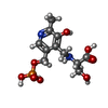  Mass: 336.235 Da / Num. of mol.: 2 / Source method: obtained synthetically / Formula: C11H17N2O8P / Feature type: SUBJECT OF INVESTIGATION Mass: 336.235 Da / Num. of mol.: 2 / Source method: obtained synthetically / Formula: C11H17N2O8P / Feature type: SUBJECT OF INVESTIGATION#4: Water | ChemComp-HOH / |  Mass: 18.015 Da / Num. of mol.: 535 / Source method: isolated from a natural source / Formula: H2O Mass: 18.015 Da / Num. of mol.: 535 / Source method: isolated from a natural source / Formula: H2OHas ligand of interest | Y | Sequence details | residue 1 is VAL according to NCBI accession WP_153841961.1 | |
|---|
-Experimental details
-Experiment
| Experiment | Method: X-RAY DIFFRACTION / Number of used crystals: 1 |
|---|
- Sample preparation
| Crystal | Density Matthews: 3.22 Å3/Da / Density % sol: 61.76 % |
|---|---|
| Crystal grow | Temperature: 298 K / Method: vapor diffusion Details: 0.1M HEPES pH 7.5, 1.4M Sodium citrate tribasic dihydrate |
-Data collection
| Diffraction | Mean temperature: 100 K / Serial crystal experiment: N |
|---|---|
| Diffraction source | Source: SYNCHROTRON / Site: SPring-8  / Beamline: BL41XU / Wavelength: 1 Å / Beamline: BL41XU / Wavelength: 1 Å |
| Detector | Type: DECTRIS EIGER X 16M / Detector: PIXEL / Date: Nov 6, 2021 |
| Radiation | Protocol: SINGLE WAVELENGTH / Monochromatic (M) / Laue (L): M / Scattering type: x-ray |
| Radiation wavelength | Wavelength: 1 Å / Relative weight: 1 |
| Reflection | Resolution: 1.9→49.35 Å / Num. obs: 92734 / % possible obs: 99.94 % / Redundancy: 26.8 % / Biso Wilson estimate: 35.59 Å2 / CC1/2: 0.999 / Rpim(I) all: 0.05079 / Rrim(I) all: 0.2614 / Net I/σ(I): 14.3 |
| Reflection shell | Resolution: 1.9→1.968 Å / Redundancy: 27.8 % / Mean I/σ(I) obs: 1 / Num. unique obs: 9141 / CC1/2: 0.652 / Rpim(I) all: 0.6137 / Rrim(I) all: 3.249 / % possible all: 99.95 |
- Processing
| Software |
| |||||||||||||||||||||||||||||||||||||||||||||||||||||||||||||||||||||||||||||||||||||||||||||||||||||||||||||||||||||||||||||||||||||||||||||||||||||||||||||||||||||||||||||||
|---|---|---|---|---|---|---|---|---|---|---|---|---|---|---|---|---|---|---|---|---|---|---|---|---|---|---|---|---|---|---|---|---|---|---|---|---|---|---|---|---|---|---|---|---|---|---|---|---|---|---|---|---|---|---|---|---|---|---|---|---|---|---|---|---|---|---|---|---|---|---|---|---|---|---|---|---|---|---|---|---|---|---|---|---|---|---|---|---|---|---|---|---|---|---|---|---|---|---|---|---|---|---|---|---|---|---|---|---|---|---|---|---|---|---|---|---|---|---|---|---|---|---|---|---|---|---|---|---|---|---|---|---|---|---|---|---|---|---|---|---|---|---|---|---|---|---|---|---|---|---|---|---|---|---|---|---|---|---|---|---|---|---|---|---|---|---|---|---|---|---|---|---|---|---|---|---|
| Refinement | Method to determine structure: FOURIER SYNTHESIS / Resolution: 1.9→49.35 Å / SU ML: 0.2723 / Cross valid method: FREE R-VALUE / σ(F): 1.36 / Phase error: 21.8558 Stereochemistry target values: GeoStd + Monomer Library + CDL v1.2
| |||||||||||||||||||||||||||||||||||||||||||||||||||||||||||||||||||||||||||||||||||||||||||||||||||||||||||||||||||||||||||||||||||||||||||||||||||||||||||||||||||||||||||||||
| Solvent computation | Shrinkage radii: 0.9 Å / VDW probe radii: 1.11 Å / Solvent model: FLAT BULK SOLVENT MODEL | |||||||||||||||||||||||||||||||||||||||||||||||||||||||||||||||||||||||||||||||||||||||||||||||||||||||||||||||||||||||||||||||||||||||||||||||||||||||||||||||||||||||||||||||
| Displacement parameters | Biso mean: 38.7 Å2 | |||||||||||||||||||||||||||||||||||||||||||||||||||||||||||||||||||||||||||||||||||||||||||||||||||||||||||||||||||||||||||||||||||||||||||||||||||||||||||||||||||||||||||||||
| Refinement step | Cycle: LAST / Resolution: 1.9→49.35 Å
| |||||||||||||||||||||||||||||||||||||||||||||||||||||||||||||||||||||||||||||||||||||||||||||||||||||||||||||||||||||||||||||||||||||||||||||||||||||||||||||||||||||||||||||||
| Refine LS restraints |
| |||||||||||||||||||||||||||||||||||||||||||||||||||||||||||||||||||||||||||||||||||||||||||||||||||||||||||||||||||||||||||||||||||||||||||||||||||||||||||||||||||||||||||||||
| LS refinement shell |
|
